

Clinical signs, magnetic resonance imaging findings and outcome in 77 cats with vestibular disease: a retrospective study
- PMID: 19932040
- PMCID: PMC11135580
- DOI: 10.1016/j.jfms.2009.10.001
Clinical signs, magnetic resonance imaging findings and outcome in 77 cats with vestibular disease: a retrospective study
Abstract
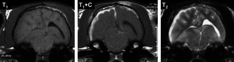
Medical records of 77 cats that had clinical signs of vestibular disease and magnetic resonance imaging (MRI) of the head were reviewed retrospectively. The aetiological, clinical and MRI characteristics were described and evaluated for a relationship with patient outcome. Forty cats (52%) had signs of central vestibular dysfunction (CVD), which was part of a multifocal disease in 17 cats (43%). The most frequent causes of CVD were inflammatory conditions (18 cats; 45%), including bacterial inflammation as an intracranial extension of otitis interna (five cats; 13%), feline infectious peritonitis (three cats; 8%) and toxoplasmosis (two cats; 5%). Neoplasia (12 cats; 30%) and vascular disease (four cats; 10%) were respectively the second and the third most frequent causes of CVD. Thiamine deficiency was diagnosed in one cat based on MRI findings and improvement following vitamin B(1) supplementation. Of 37 cats (48%) with peripheral vestibular dysfunction (PVD), idiopathic vestibular syndrome (IVS) was suspected in 16 (43%) and otitis media/interna was suspected in 16 (43%). Within the group of cats with evident MRI lesions, the location of the imaged lesions agreed with the clinical classification of vestibular dysfunction in 52/55 (95%) cats. Most of the cats (nine cases; 56%) with presumed IVS had rapid and complete recovery of their clinical signs. As most of these cats presented with progressive clinical signs over 3 weeks they were classified as having 'atypical' IVS to differentiate them from cats with the typical non-progressive IVS. No underlying systemic diseases were documented in any of these cases. Statistically significant predictors of survival included neurolocalisation (central or peripheral vestibular system), age and gender. No difference in survival was observed between cats with presumed idiopathic peripheral syndrome and cats with otitis media/interna.
Copyright 2009 ISFM and AAFP. All rights reserved.
Figures


Similar articles
-
Clinical reasoning in feline vestibular syndrome: which presenting features are the most important?J Feline Med Surg. 2021 Aug;23(8):669-678. doi: 10.1177/1098612X20970869. Epub 2020 Nov 12. J Feline Med Surg. 2021. PMID: 33176542 Free PMC article.
-
Clinical signs, MRI findings and outcome in dogs with peripheral vestibular disease: a retrospective study.BMC Vet Res. 2020 May 25;16(1):159. doi: 10.1186/s12917-020-02366-8. BMC Vet Res. 2020. PMID: 32450859 Free PMC article.
-
Vestibular disease: diseases causing vestibular signs.Compend Contin Educ Vet. 2012 Jul;34(7):E2. Compend Contin Educ Vet. 2012. PMID: 22847321 Review.
-
Concurrent idiopathic vestibular syndrome and facial nerve paralysis in a cat.Aust Vet J. 2015 Jul;93(7):252-4. doi: 10.1111/avj.12338. Aust Vet J. 2015. PMID: 26113351
-
The neurology of balance: function and dysfunction of the vestibular system in dogs and cats.Vet J. 2010 Sep;185(3):247-58. doi: 10.1016/j.tvjl.2009.10.029. Epub 2009 Nov 26. Vet J. 2010. PMID: 19944632 Review.
Cited by
-
Clinical reasoning in feline vestibular syndrome: which presenting features are the most important?J Feline Med Surg. 2021 Aug;23(8):669-678. doi: 10.1177/1098612X20970869. Epub 2020 Nov 12. J Feline Med Surg. 2021. PMID: 33176542 Free PMC article.
-
Feline neurological diseases in a veterinary neurology referral hospital population in Japan.J Vet Med Sci. 2019 Jun 21;81(6):879-885. doi: 10.1292/jvms.18-0447. Epub 2019 Apr 30. J Vet Med Sci. 2019. PMID: 31061248 Free PMC article.
-
A Pandora's box in feline medicine: presenting signs and surgical outcomes in 58 previously hoarded cats with chronic otitis media-interna.J Feline Med Surg. 2023 Sep;25(9):1098612X231197089. doi: 10.1177/1098612X231197089. J Feline Med Surg. 2023. PMID: 37728478 Free PMC article.
-
Magnetic Resonance Imaging Findings in 13 Neurologic Pot-Bellied Pigs.Front Vet Sci. 2020 Jan 31;7:21. doi: 10.3389/fvets.2020.00021. eCollection 2020. Front Vet Sci. 2020. PMID: 32076601 Free PMC article.
-
Utility of a Modified Penlight-Cover Test for Neurolocalization of Lesions Based on Visual Suppression of Nystagmus in Dogs and Cats With Vestibular Disease.J Vet Intern Med. 2025 Jul-Aug;39(4):e70182. doi: 10.1111/jvim.70182. J Vet Intern Med. 2025. PMID: 40577055 Free PMC article.
References
-
- Kornegay J.N. Ataxia, head tilt, nystagmus. Vestibular diseases, Probl Vet Med 3, 1991, 417–425. - PubMed
-
- Muñana K.R. Head tilt and nystagmus. Platt S.R., Olby N.J. BSAVA manual of canine and feline neurology, 3rd edn, 2004, British Small Animal Veterinary Association: Gloucester, 155–171.
-
- Dickinson P.J., Keel M.K., Higgins R.J., et al. Clinical and pathologic features of oligodendrogliomas in two cats, Vet Pathol 37, 2000, 160–167. - PubMed
-
- Garosi L.S., Dennis R., Penderis J., et al. Results of magnetic resonance imaging in dogs with vestibular disorders: 85 cases (1996–1999), J Am Vet Med Assoc 128, 2001, 385–391. - PubMed
MeSH terms
LinkOut - more resources
Full Text Sources
Medical
Research Materials
Miscellaneous
