Review
. 2010 May 1;341(1):66-83.
doi: 10.1016/j.ydbio.2009.10.024. Epub 2009 Nov 20.

Myoblast fusion: when it takes more to make one

Affiliations
Review

Myoblast fusion: when it takes more to make one

Kate Rochlin et al. Dev Biol. .

Abstract

Cell-cell fusion is a crucial and highly regulated event in the genesis of both form and function of many tissues. One particular type of cell fusion, myoblast fusion, is a key cellular process that shapes the formation and repair of muscle. Despite its importance for human health, the mechanisms underlying this process are still not well understood. The purpose of this review is to highlight the recent literature pertaining to myoblast fusion and to focus on a comparison of these studies across several model systems, particularly the fly, zebrafish and mouse. Advances in technical analysis and imaging have allowed identification of new fusion genes and propelled further characterization of previously identified genes in each of these systems. Among the cellular steps identified as critical for myoblast fusion are migration, recognition, adhesion, membrane alignment and membrane pore formation and resolution. Importantly, striking new evidence indicates that orthologous genes govern several of these steps across these species. Taken together, comparisons across three model systems are illuminating a once elusive process, providing exciting new insights and a useful framework of genes and mechanisms.


Figures

Figure 1
Figure 1. Myofibers in a variety of model systems form from the fusion of mononucleated muscle precursors
(A-D) Nuclei were visualized using a nuclear DsRed transgene (red) (A), DAPI (blue) (B, C) or hematoxylin (purple) (D). (A) Four hemisegments of a Drosophila embryo were analyzed by immunohistochemistry using antibodies against tropomyosin (green). (B) The syncytial fast-twitch muscle fibers of a wild-type zebrafish embryo were labeled with antibodies against fast myosin light chain (red). Two fast muscle fibers are highlighted by GFP expression (green) from a skeletal muscle actin∷gfp transgene. (C) C2C12 myoblasts, a satellite cell-derived mouse myoblast cultured cell line, were analyzed by immunohistochemistry using antibodies against myosin heavy chain (green). The scale bar represents 40 μm. (D) Cross sections of a major leg muscle, the gastrocnemius, of an adult mouse stained with hematoxylin and eosin. Note that the nuclei are peripherally located and, unlike in other panels, the multinucleate nature of the myofiber is not clearly evident from this section.
Figure 2
Figure 2. Cellular and subcellular behaviors that occur during myoblast fusion
Several cellular steps occur during myoblast fusion; each step coincides with a defined series of subcellular events. (A) Cells first migrate toward their fusing partner, concurrently with actin polymerization (red lines) and expression of transmembrane attractants (small rectangles) to guide the migrating cell. (B) The cells then touch and adhere, leading to localization of the cell type-specific transmembrane proteins. (C) This leads to an accumulation of actin (red oval), and the formation of the FuRMAS (purple ring) at the site of fusion. Subsequently, a number of fusion proteins known collectively as the “fusion machinery” are localized to the site of fusion, presumably through vesicular trafficking. The area outlined by the grey box is examined in more detail in Figure 3. (D) This is followed by membrane breakdown and the removal of vesiculating membrane and the fusion machinery components. (E) Finally, the cell must reset for the next round of fusion by expressing appropriate levels of transmembrane attractant. This process will repeat iteratively until the final muscle or fiber size is achieved.
Figure 3
Figure 3. Current working model of the genes required for myoblast fusion in Drosophila
A simplified model that has been updated to reflect recently identified fusion genes and to illustrate the conservation of proteins to the zebrafish and mouse myoblast fusion paradigms. Note that nuclei are in white, myotube in is grey and the FCM is in blue. The actin focus and FuRMAS are depicted as a red oval and a purple ring, respectively. Rectangles represent proteins that have a conserved role in myoblast fusion in multiple systems. Ovals represent proteins with known homologs in vertebrates that have no role in fusion described to date. Diamonds represent proteins that have no known homologs in vertebrates. Solid arrows denote well-characterized biochemical interactions, dashed arrows indicate genetic and/or suggested biochemical interactions and white arrows designate interactions that are suggested from work on orthologous proteins in other contexts. While this depiction suggests specific interacting partners for each transmembrane protein, there is evidence that this may not be the case as interactions between Duf and Hbs have been shown to mediate cell adhesion in vitro. Additionally, the reader is cautioned that, although strong biochemical exists for Mbc-mediated Rac activation and for the Rac→ SCAR complex→ Arp2/3 pathway, there is not yet evidence for a complete pathway linking Mbc to Arp2/3-dependent actin polymerization.
Figure 4
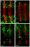
Figure 4. kette mutant FCMs can migrate towards ectopically expressed Dumbfounded (Duf), which acts as an FCM attractant in vivo
(A) wild type, (B) kette mutant, (C) Wingless (Wg)-GAL4 > UAS-Duf and (D) Wg-GAL4 > UAS-Duf; kette embryos. Myofibers and FCMs are in green; Wg epidermal cells, the source of the attractant Duf in the experimental embryos are in red. In wild type (A) and kette mutant (B) embryos, no myoblasts are present in the ventral ectoderm. However, when the expression of Duf is driven by Wg GAL4 in wild-type embryos (C), FCMs migrate ectopically into the ventral ectoderm. When Duf is expressed in kette mutant embryos (D), FCMs are capable of migrating towards the source of the attractant. The scale bar represents 20 μm.
Figure 5
Figure 5. The activities of Rac1 and Kirrel/Nephrin are required for vertebrate myoblast fusion
(A-C) The zebrafish fast-twitch syncytium in a control embryo (A), an embryo expressing constitutively active Rac1 (caRac1) (B) and a kirrel morphant zebrafish embryo (C) were analyzed by immunohistochemistry using antibodies against myosin heavy chain (red) and hemagglutinin to label Rac1 (green) in (B). (A) The fast-twitch muscle fibers of a wild-type zebrafish embryo contain multiple nuclei. (B) The fast-twitch syncytium in zebrafish embryos expressing caRac1 are hyperfused, containing more nuclei than in wild-type syncytium. (C) In kirrel morphants, there are large numbers of unfused fast-twitch precursors. (D-E) Longitudinal sections of myofibers from the proximal forelimb of control (D) and conditional Rac1 mutant (E) mice at E13.5 were analyzed by immunohistochemistry using antibodies against desmin (green) and MyoD (red). The scale bar represents 50 μm. (E) Myofibers in conditional Rac1 mutant mice (E) are short and thin, indicative of a fusion impairment in vivo, in comparison to control myofibers, which form long, multinucleated fibers (D). (F-G) Myoblasts isolated from control (F) and nephrin null (G) neonatal mice were analyzed by immunohistochemistry using antibodies against desmin (red). (F) Control myocytes form large, multinucleated cells after 4 days of differentiation in vitro. (G) Myocytes from nephrin null mice differentiate, but fail to fuse during the same time period. Nuclei were visualized with DAPI (blue) (A-C, F, G) or MyoD (D, E).
Figure 6
Figure 6. Venn diagram depicting a subset of genes that have been analyzed in each model system
The model systems Drosophila, mouse and zebrafish are illustrated as blue, red and yellow partially overlapping myoblasts. A subset of genes analyzed to date are listed in each myoblast. Genes unique to a model organism are located in non-overlapping, monochromatic regions of the suitable myoblast. Genes that have been studied in multiple models are localized to the appropriate overlapping regions. Genes that have been analyzed in more than one system are phenotypically compared in Table 3.

Similar articles

Cited by

References

    1. Artero RD, Castanon I, Baylies MK. The immunoglobulin-like protein Hibris functions as a dose-dependent regulator of myoblast fusion and is differentially controlled by Ras and Notch signaling. Development. 2001;128:4251–64. - PubMed
    1. Bae GU, Gaio U, Yang YJ, Lee HJ, Kang JS, Krauss RS. Regulation of myoblast motility and fusion by the CXCR4-associated sialomucin, CD164. J Biol Chem. 2008;283:8301–9. - PMC - PubMed
    1. Balagopalan L, Chen MH, Geisbrecht ER, Abmayr SM. The CDM superfamily protein MBC directs myoblast fusion through a mechanism that requires phosphatidylinositol 3,4,5-triphosphate binding but is independent of direct interaction with DCrk. Mol Cell Biol. 2006;26:9442–55. - PMC - PubMed
    1. Barletta GM, Kovari IA, Verma RK, Kerjaschki D, Holzman LB. Nephrin and Neph1 co-localize at the podocyte foot process intercellular junction and form cis hetero-oligomers. J Biol Chem. 2003;278:19266–71. - PubMed
    1. Bate M. The embryonic development of larval muscles in Drosophila. Development. 1990;110:791–804. - PubMed

Publication types

LinkOut - more resources