. 2010 Jan;18(1):88-96.
doi: 10.1016/j.joca.2009.11.006. Epub 2009 Nov 18.

Phenotypic characterization of epiphycan-deficient and epiphycan/biglycan double-deficient mice

Affiliations

Phenotypic characterization of epiphycan-deficient and epiphycan/biglycan double-deficient mice

S Nuka et al. Osteoarthritis Cartilage. 2010 Jan.

Abstract

Objective: To characterize the in vivo role epiphycan (Epn) has in cartilage development and/or maintenance.

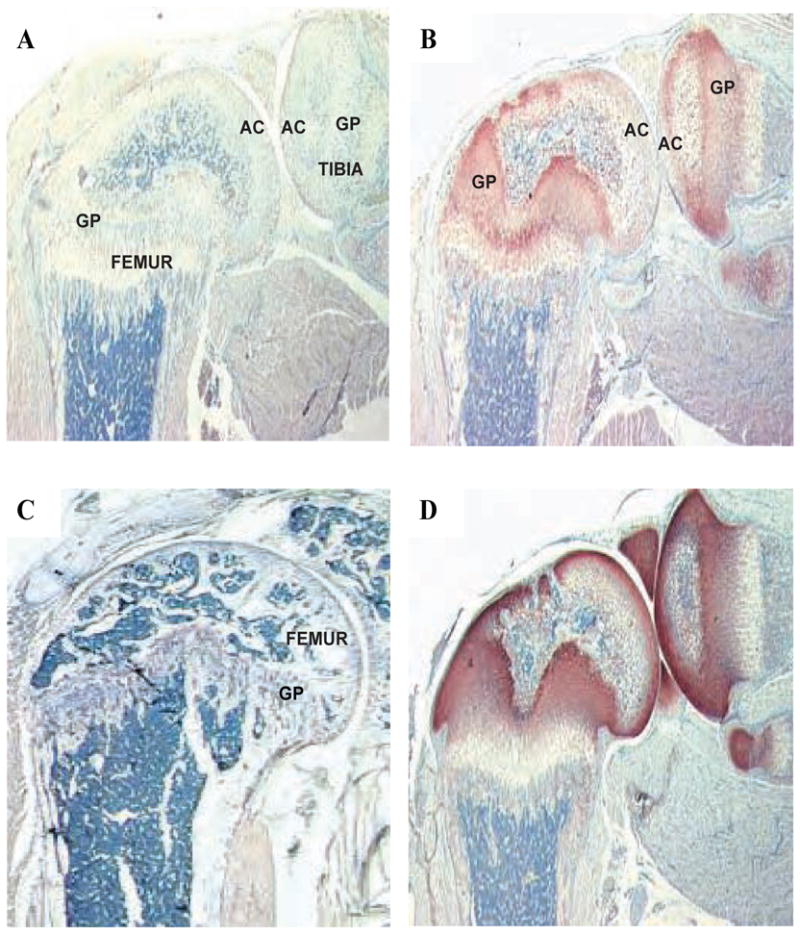
Methods: Epn-deficient mice were generated by disrupting the Epn gene in mouse embryonic stem cells. Epn/biglycan (Bgn) double-deficient mice were produced by crossing Epn-deficient mice with Bgn-deficient mice. Whole knee joint histological sections were stained using van Gieson or Fast green/Safranin-O to analyze collagen or proteoglycan content, respectively. Microarray analysis was performed to detect gene expression changes within knee joints.

Results: Epn-deficient and Epn/Bgn double-deficient mice appeared normal at birth. No significant difference in body weight or femur length was detected in any animal at 1 month of age. However, 9-month Epn/Bgn double-deficient mice were significantly lighter and had shorter femurs than wild type mice, regardless of gender. Male Epn-deficient mice also had significantly shorter femurs than wild type mice at 9 months. Most of the deficient animals developed osteoarthritis (OA) with age; the onset of OA was observed earliest in Epn/Bgn double-deficient mice. Message RNA isolated from Epn/Bgn double-deficient knee joints displayed increased matrix protein expression compared with wild type mice, including other small leucine-rich proteoglycan (SLRP) members such as asporin, fibromodulin and lumican.

Conclusion: Similar to other previously studied SLRPs, EPN plays an important role in maintaining joint integrity. However, the severity of the OA phenotype in the Epn/Bgn double-deficient mouse suggests a synergy between these two proteins. These data are the first to show a genetic interaction involving class I and class III SLRPs in vivo.


Conflict of interest statement

CONFLICT OF INTEREST STATEMENT

No conflicts of interests are associated with this manuscript.

Figures

Figure 1
Figure 1. Generation of Epn-deficient mice
A, At the top is a schematic representation of the partial Epn gene locus; in the middle is the targeting vector created to disrupt the Epn gene; at the bottom is the final product with the disrupted Epn allele. B, Mouse ES cell genomic DNA digested with ScaI and subjected to Southern blot analysis. The radiolabeled 5’ probe detects an 8 kb fragment from the wild type Epn allele and a 14 kb fragment from the interrupted Epn allele. Lane 1 is an untargeted ES clone and lanes 2-7 are targeted ES clones. C, Mouse ES cell genomic DNA digested with BglII and subjected to Southern blot analysis. The radiolabeled 3’ probe detects a 4.8 kb fragment from the wild type Epn allele and an 11.4 kb fragment from the interrupted Epn allele. Lane 1 is an untargeted ES clone and lanes 2-7 are targeted ES clones.D, Genotyping of the mice. A 2.6 kb fragment is amplified from the wild type Epn allele with the primer pair Epnlfw/Epn2rv (lanes 3 and 5), whereas a 0.4 kb fragment is amplified from the interrupted Epn allele with the primer pair Bpa/Epn2rv (lanes 2 and 4).
Figure 2
Figure 2. Immunohistological analysis of murine knee joints using the anti- Epn antibody, R561, or the anti-Bgn antibody, LF-159
Panels A, B, and D are oriented in the same manner, with the femur on the left and the tibia on the right. A, R561 antibody staining of a 2-week-old Epn-deficient tibia-femoral joint. AC = articular cartilage and GP = growth plate. B, R561 antibody staining of a 2- week-old wild type tibia-femoral joint. Epn is detected in the growth plate and articular cartilage. C, R561 antibody staining of a 2-month-old wild type femur showing very weak expression of Epn protein in the growth plate. D, LF-159 antibody staining of a 2-week-old wild type tibia-femoral joint. Bgn is detected in the growth plate, articular cartilage, and the meniscus.
Figure 3
Figure 3. Mean body weights of wild type and SLRP-deficient mice at 1 and 9 months of age
The asterisks denote a significant difference in body weight as compared to wild type mice (p<0.001). The error bars denote the SEM for each genotype.
Figure 4
Figure 4. Safranin O staining of the tibia-femoral joint in 9 month old mice
In each panel, the femur is at the top and the tibia is at the bottom of the picture. A, Representative joint section from a wild type animal. B, Representative joint section from an Epn-deficient mouse. C, Representative joint section from a Bgn-deficient mouse. D, Representative joint section from an Epn/Bgn double-deficient mouse. The asterisks in panels B, C, and D denote fibrillation as well as articular cartilage and proteoglycan loss.
Figure 5
Figure 5. Mean scores of OA severity in male wild type and SLRP-deficient mice
Sections of the tibia-femoral joint in wild-type, Epn-deficient, Bgn-deficient, and Epn/Bgn double-deficient mice were scored for OA using the criteria of Maier and Wilhelmi (30). A minimum of 4 mice were examined at every age for each genotype. The age of the mice in months is given on the x-axis, while the OA grade is shown on the y-axis. The error bars denote the SEM for each genotype. * p≤0.053, ** p = 0.036, and *** p < 0.005 compared to wild type mice at that age. With Bonferroni correction, a p value of ≤0.02 is necessary to reach statistical significance.
Figure 6
Figure 6. Collagen analysis in tibia-femoral joints
Sections of 3 month old male mouse tibia-femoral joints from wild type, Bgn-deficient, and Epn/Bgn double-deficient mice were stained with van Giesen to analyze collagen content. The image at the top left of each panel is a 6-fold magnified image of the area denoted by the black box (n = 3).

Similar articles

Cited by

References

    1. Johnson J, Shinomura T, Eberspaecher H, Pinero G, Decrombrugghe B, Hook M. Expression and localization of PG-Lb/epiphycan during mouse development. Dev Dyn. 1999;216:499–510. - PubMed
    1. Roughley PJ. The structure and function of cartilage proteoglycans. Eur Cell Mater. 2006;12:92–101. - PubMed
    1. Schmidt G, Robenek H, Harrach B, Glossl J, Nolte V, Hormann H, et al. Interaction of small dermatan sulfate proteoglycan from fibroblasts with fibronectin. J Cell Biol. 1987;104:1683–1691. - PMC - PubMed
    1. Winnemoller M, Schon P, Vischer P, Kresse H. Interactions between thrombospondin and the small proteoglycan decorin: interference with cell attachment. Eur J Cell Biol. 1992;59:47–55. - PubMed
    1. Dugan TA, Yang VW, McQuillan DJ, Hook M. Decorin modulates fibrin assembly and structure. J Biol Chem. 2006 - PubMed

Publication types