

Three-dimensional echocardiographic assessment of changes in mitral valve geometry after valve repair
- PMID: 19932245
- PMCID: PMC3019306
- DOI: 10.1016/j.athoracsur.2009.07.007
Three-dimensional echocardiographic assessment of changes in mitral valve geometry after valve repair
Abstract
Background: Application of annuloplasty rings during mitral valve (MV) repair has been shown to significantly change the mitral annular geometry. Until recently, a comprehensive two-dimensional echocardiographic evaluation of annular geometric changes was difficult owing to its nonplanar orientation. In this study, an analysis of the three-dimensional intraoperative transesophageal echocardiographic evaluation of the MV annulus is presented before and immediately after repair.
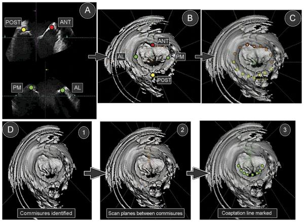
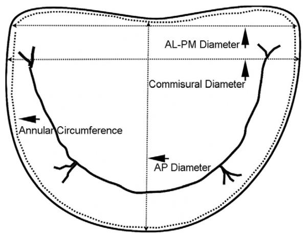
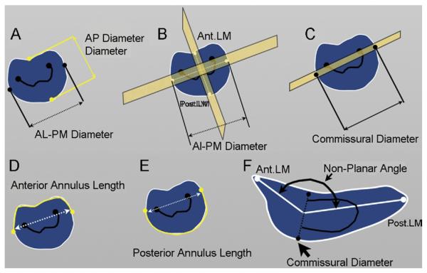
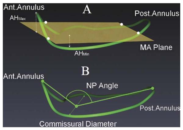
Methods: We performed three-dimensional geometric analysis on 75 patients undergoing MV repair during coronary artery bypass graft surgery for mitral regurgitation or myxomatous mitral valve disease. Geometric analysis of the MV was performed before and immediately after valve repair with full rings and annuloplasty bands. The acquired three-dimensional volumetric data were analyzed in the operating room. Specific measurements included annular diameter, leaflet lengths, the nonplanarity angle, and the circularity index. Before and after repair data were compared.
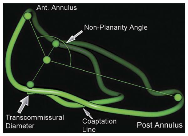
Results: Complete echocardiographic assessment of the MV was feasible in 69 of 75 patients (92%) within 2 to 3 minutes of acquisition. Placement of full rings resulted in an increase in the nonplanarity angle or a less saddle shape of the native mitral annulus (137 +/- 14 versus 146 +/- 14; p = 0.002. By contrast, the nonplanarity angle did not change significantly after placement of partial rings.
Conclusions: Mitral annular nonplanarity can be assessed in the operating room. Application of full annuloplasty rings resulted in the mitral annulus becoming more planar. Partial annuloplasty bands did not significantly change the nonplanarity angle. Neither of the two types of rings restored the native annular planarity.
Figures
References
-
- Fedak PW, McCarthy PM, Bonow RO. Evolving concepts and technologies in mitral valve repair. Circulation. 2008;117:963–74. - PubMed
-
- Flachskampf FA, Chandra S, Gaddipatti A, et al. Analysis of shape and motion of the mitral annulus in subjects with and without cardiomyopathy by echocardiographic 3-dimensional reconstruction. J Am Soc Echocardiogr. 2000;13:277–87. - PubMed
-
- Gillinov AM, Cosgrove DM, Shiota T, et al. Cosgrove-Edwards annuloplasty system: midterm results. Ann Thorac Surg. 2000;69:717–21. - PubMed
-
- Glasson JR, Komeda M, Daughters GT, Bolger AF, Ingels NB, Miller DC. Loss of three-dimensional canine mitral annular systolic contraction with reduced left ventricular volumes. Circulation. 1996;94:II152–8. - PubMed
-
- Glasson JR, Komeda M, Daughters GT, et al. Three-dimensional dynamics of the canine mitral annulus during ischemic mitral regurgitation. Ann Thorac Surg. 1996;62:1059–68. - PubMed
Publication types
MeSH terms
Grants and funding
LinkOut - more resources
Full Text Sources
Medical
