. 2009 Dec 4;326(5958):1373-9.
doi: 10.1126/science.1181829. Epub 2009 Oct 22.

Structural mechanism of abscisic acid binding and signaling by dimeric PYR1

Affiliations

Structural mechanism of abscisic acid binding and signaling by dimeric PYR1

Noriyuki Nishimura et al. Science. .

Abstract

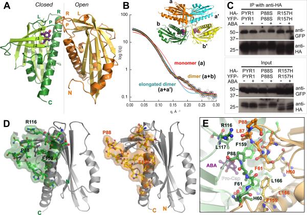
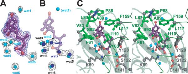
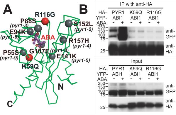
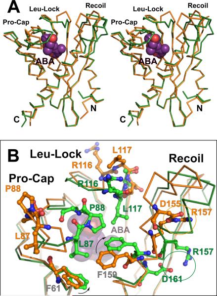
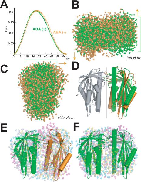
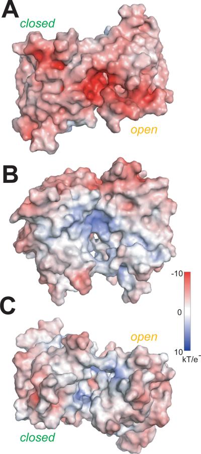
The phytohormone abscisic acid (ABA) acts in seed dormancy, plant development, drought tolerance, and adaptive responses to environmental stresses. Structural mechanisms mediating ABA receptor recognition and signaling remain unknown but are essential for understanding and manipulating abiotic stress resistance. Here, we report structures of pyrabactin resistance 1 (PYR1), a prototypical PYR/PYR1-like (PYL)/regulatory component of ABA receptor (RCAR) protein that functions in early ABA signaling. The crystallographic structure reveals an alpha/beta helix-grip fold and homodimeric assembly, verified in vivo by coimmunoprecipitation. ABA binding within a large internal cavity switches structural motifs distinguishing ABA-free "open-lid" from ABA-bound "closed-lid" conformations. Small-angle x-ray scattering suggests that ABA signals by converting PYR1 to a more compact, symmetric closed-lid dimer. Site-directed PYR1 mutants designed to disrupt hormone binding lose ABA-triggered interactions with type 2C protein phosphatase partners in planta.


Figures

Fig. 1
Fig. 1
Dimeric structure of ABA sensor PYR1. (A) Crystallographic asymmetric dimer shown as ribbons (labeled β-strands and helices) with ABA (purple ball-and-stick model with red oxygen atoms) bound beneath the closed lid of one subunit (left), in a large cavity between the β-sheet and long C-terminal α-helix. Labeled Pro-Cap (P), Leu-Lock (L) and Recoil (R) structural motifs undergo ABA-induced conformational changes. Vertical line (center) indicates pseudo 2-fold axis relating the subunits. (B) Theoretical SAXS curve (orange line) for asymmetric crystallographic dimer (a+b in inset) matches experimental SAXS data for PYR1 without ABA (open circles), whereas curves calculated assuming a monomer (red) or elongated a+a' dimer (blue) from crystal lattice (see inset) do not. (C) Co-immunoprecipitation from extracts of plant leaves expressing YFP-tagged PYR1 and HA-tagged PYR1 without (−) and with (+) exogenously applied ABA confirm dimeric PYR1 assembly. After co-immunoprecipitation using an anti-HA matrix, immunoprecipitated (above) and input (below) samples were detected with anti-GFP and anti-HA antibodies. PYR1 wild-type and mutants P88S and R157H are homodimeric in planta, as shown by anti-GFP antibody labeling of YFP-tagged PYR1 co-immunoprecipitated with HA-tagged PYR1. (D) Residues and buried surface area contributed to dimer interface by closed-lid (left) and open-lid (right) subunit conformations in asymmetric dimer. (E) PYR1 dimer interface viewed looking down from top in (A) at the interacting lids: open (orange) and closed (green) over ABA (purple). Dimer contacts include the interacting Pro-Cap structural motifs (foreground), plus a side-chain-to-main-chain hydrogen bond from Arg116 in the ABA-bound subunit (top left) to Leu87 in the ABA-free subunit.
Fig. 2
Fig. 2
Water-filled ABA-binding cavity. (A) ABA (purple ball and stick model, with red oxygen atoms) and adjacent, ordered, water molecules (cyan spheres) inside the PYR1 cavity, shown with electron density (mesh). Omit Fo-Fc density for ABA contoured at 3σ (blue) and 4σ (magenta); 2Fo-Fc electron density for water molecules contoured at 1σ (black). All maps were calculated after “shaking” coordinates to reduce phase bias. (B) Ordered water molecules (blue spheres) within the ABA-free subunit cavity, shown with associated 2Fo-Fc electron density, as in (A). ABA (purple) and water molecules (cyan) from ABA-bound PYR1 subunit (shown in A) are superimposed showing conserved, water positions. ABA displaces one water molecule (wat7) with the carboxylate, shifts a second (wat2), and introduces or stabilizes a third (wat1), which interacts with the ABA carbonyl to stabilize lid closure. (C) Stereo view of PYR1 residues contributing to the ABA binding site. Hydrophobic side chains (green ball-and-stick) surround the ABA ring, whereas hydrogen-bonded (red dashed lines) internal water molecules (cyan spheres) link ABA oxygen atoms (red) to PYR1 hydrophilic side chains (gray ball-and-stick with red oxygen and blue nitrogen atoms) projecting into the binding cavity. Larger gray spheres show Cα atoms. Lys59, Phe61, Arg79, Val83, Leu87, Pro88, Ala89, Ser92, Glu94, Ile110, Leu117, Tyr120, Ser122, Glu141, Phe159, Val163 and Asn167 contribute to forming this large internal cavity.
Fig. 3
Fig. 3
Disruption of PYR1-ABI1 interactions by single-site PYR1 mutations. (A) PYR1 mutants designed from the structure (red) and identified after chemical mutagenesis and screening (gray) (6) are mapped to the PYR1 subunit structure (green Cα trace). (B) Co-immunoprecipitation from extracts of plant leaves expressing YFP-tagged ABI1 phosphatase with HA-tagged wild-type and mutant PYR1 proteins in planta, both in the absence (−) and presence (+) of exogenously applied ABA. After co-immunoprecipitation by an anti-HA matrix, immunoprecipitated (top) and input (bottom) samples were detected with anti GFP and anti-HA antibodies (labeled at right). Structure-based PYR1 mutants designed to disrupt ABA binding (K59Q and R116G) folded properly (fig. S5), but lost ABA-induced interactions with ABI1 phosphatase.
Fig. 4
Fig. 4
ABA-induced subunit conformational changes. (A) Stereo image showing superposition of ABA-free (orange) and ABA-bound (green) PYR1 Cα traces. ABA-induced helix coiling by the Recoil motif (upper right) is coupled to lid closure over ABA (purple with red oxygen atoms) by the Pro-Cap and Leu-Lock structural motifs (upper left). (B) Enlarged view of ABA-triggered conformational changes in these three structural motifs that close the lid over bound ABA, colored as in (A). ABA (beneath center) triggers rotation of Phe159 (arrow) to coil the Recoil motif into helix α3 (diagonal at right), switching the Arg157 charge-charge interaction (circled) to a new partner within (rather than outside) this helix. Pro88 isomerization from trans (orange, far left) to cis (green, center) converts the open-lid Pro-Cap to the closed-lid conformation, clamping Leu87, Pro88 and Ala89 over ABA. Leu117 (orange, top center) locks down (green, center) against ABA, closing the Leu-Lock, and flipping the preceding Arg116 side chain (orange, top center) toward the opposing subunit (forward and slightly to the right in this view) across the dimer interface (see also Fig. 1E).
Fig. 5
Fig. 5
ABA-induced changes in dimer assembly analyzed by SAXS. (A) The pair distance distribution function describing intramolecular distances in the PYR1 dimer in the absence of ABA (orange) becomes narrower and shifts to shorter distances in the presence of saturating ABA (green). (B, C) Two sets of eight independent ab initio bead models for the PYR1 dimer, representing SAXS experimental data in the absence (orange) or presence (green) of saturating ABA. Green brackets mark flatter PYR1 disk in the presence of saturating ABA. Orange arrows indicate greater thickness and asymmetry of PYR1 dimer in absence of ABA. ABA-induced changes in subunit orientation make the PYR1 dimer disk flatter and more compact, as seen from top (B) and side (C) relative to orientation in D. (D) Cartoons (depicting α-helices as cylinders and β-strands as arrows) of the asymmetric crystallographic dimer and a symmetric closed-lid dimer model, aligned by superposition of their common subunit (gray). The ~10° difference in orientation between the second subunits of each dimer (right), highlights the differences between the pseudo 2-fold axis (~170°) relating subunits (gray and orange) of the asymmetric crystallographic dimer and the exact 2-fold axis (vertical line) relating subunits (gray and green) of the symmetric dimer model. (E, F) Independently determined bead models (four sets of colored dots) representing SAXS results in the absence (E) and presence (F) of ABA, each aligned with the corresponding PYR1 structural model (D). The PYR1 dimer assembly shapes determined by SAXS show excellent fits to the crystallographic asymmetric dimer (E) and symmetric dimer model (F). The biconcave, red blood cell shape of the PYR1 dimer is seen by decreased bead density in the center of the PYR1 disks, as well as in cross section (B), particularly with saturating ABA.
Fig. 6
Fig. 6
PYR1 molecular surface color-coded by electrostatic potential. ABA-induced changes in subunit orientation produce conformational changes in the disk-shaped PYR1 dimer at the (A) interacting lids (top), (B) concave sides, aligned as in Fig. 5 (D and E), and (C) the cleft (bottom) between C-terminal helices. Conformational changes of charged residues in the Leu-Lock (Glu114 and Arg116) and Recoil (Glu149, Glu153, Asp154, Asp155 and Arg157) motifs of each subunit reduce electrostatic surface potential upon lid closure (A).

Comment in

References

    1. Ohkuma K, Lyon JL, Addicott FT, Smith OE. Science. 1963;142:1592. - PubMed
    1. Eagles CF, Wareing PF. Nature. 1963;199:874.
    1. Finkelstein RR, Gampala SS, Rock CD. Plant Cell. 2002;14:S15. - PMC - PubMed
    1. Yamaguchi-Shinozaki K, Shinozaki K. Annu Rev Plant Biol. 2006;57:781. - PubMed
    1. Ma Y, et al. Science. 2009;324:1064. - PubMed

Publication types

MeSH terms

Associated data