

Ultrahigh-resolution measurement by optical coherence tomography of dynamic tear film changes on contact lenses
- PMID: 19933178
- PMCID: PMC2868399
- DOI: 10.1167/iovs.09-4389
Ultrahigh-resolution measurement by optical coherence tomography of dynamic tear film changes on contact lenses
Abstract
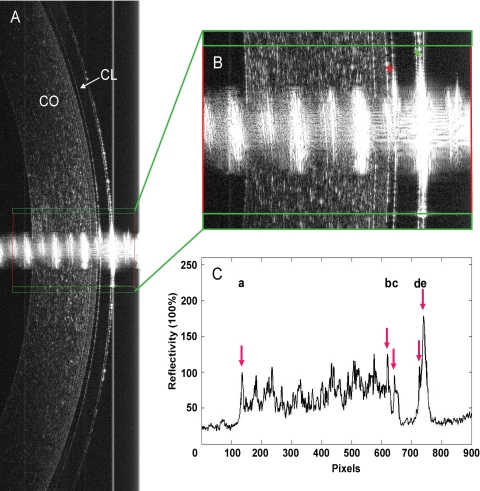
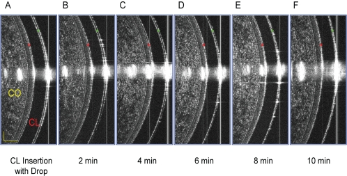
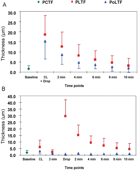
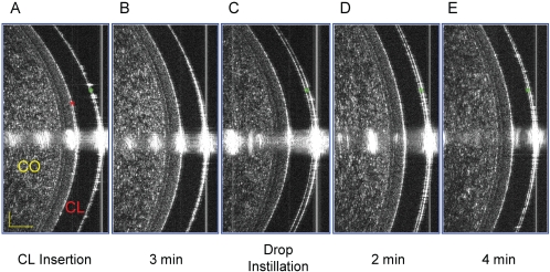
Purpose. To determine the dynamic pre- and postlens tear film (PLTF and PoLTF) thicknesses by using optical coherence tomography (OCT). Methods. Ultrahigh-resolution OCT was used to image the tear film of 22 subjects before and after contact lens wear. A soft lens with 1 drop of artificial tears on its concave surface was inserted onto one randomly selected eye. OCT images were taken before insertion, immediately afterward, and every 2 minutes for 10 minutes. For the contralateral eye, the lens inserted was not prewetted on the concave surface. OCT images were taken before insertion, immediately afterward, and at 3 minutes. Then another drop was instilled, and images were taken immediately afterward and every 2 minutes for 10 minutes. Images were processed by custom software to yield tear film thickness. Results. The thickness of precorneal tear film (PCTF) was 1.9 +/- 0.9 mum. The PoLTF was visualized clearly in all cases immediately after lens insertion, with 1 drop on the lens concave surface. Through the first 6 minutes after insertion, the PoLTF was greater than the PCTF. The PLTF (n = 12) and PoLTF (n = 9) were visualized immediately after lens insertion. After 3 minutes, the PLTF in most subjects and PoLTF in all subjects were invisible. The thickness of the PLTF increased after the instillation of artificial tears, whereas the PoLTF did not at any checkpoint for 10 minutes. Conclusions. Ultrahigh-resolution OCT is a promising tool for measuring the tear film. The PoLTF did not increase after instillation of artificial tears.
Figures
Similar articles
-
Diquafosol Ophthalmic Solution Increases Pre- and Postlens Tear Film During Contact Lens Wear in Rabbit Eyes.Eye Contact Lens. 2017 Nov;43(6):378-382. doi: 10.1097/ICL.0000000000000283. Eye Contact Lens. 2017. PMID: 27243352
-
Precorneal and pre- and postlens tear film thickness measured indirectly with optical coherence tomography.Invest Ophthalmol Vis Sci. 2003 Jun;44(6):2524-8. doi: 10.1167/iovs.02-0731. Invest Ophthalmol Vis Sci. 2003. PMID: 12766052 Clinical Trial.
-
The impact of contact lens care solutions on the thickness of the tear film and contact lens.Cornea. 2005 Oct;24(7):825-32. doi: 10.1097/01.ico.0000157904.52305.29. Cornea. 2005. PMID: 16160499 Clinical Trial.
-
Protection against corneal hyperosmolarity with soft-contact-lens wear.Prog Retin Eye Res. 2022 Mar;87:101012. doi: 10.1016/j.preteyeres.2021.101012. Epub 2021 Sep 29. Prog Retin Eye Res. 2022. PMID: 34597771 Review.
-
Central Corneal Edema with Scleral-Lens Wear.Curr Eye Res. 2018 Nov;43(11):1305-1315. doi: 10.1080/02713683.2018.1500610. Epub 2018 Aug 6. Curr Eye Res. 2018. PMID: 30015536 Review.
Cited by
-
Ocular Surface Changes Associated with Ophthalmic Surgery.J Clin Med. 2021 Apr 12;10(8):1642. doi: 10.3390/jcm10081642. J Clin Med. 2021. PMID: 33921534 Free PMC article. Review.
-
Measurement of a multi-layered tear film phantom using optical coherence tomography and statistical decision theory.Biomed Opt Express. 2014 Nov 24;5(12):4374-86. doi: 10.1364/BOE.5.004374. eCollection 2014 Dec 1. Biomed Opt Express. 2014. PMID: 25574445 Free PMC article.
-
Applications of Anterior Segment Optical Coherence Tomography in Cornea and Ocular Surface Diseases.J Ophthalmol. 2016;2016:4971572. doi: 10.1155/2016/4971572. Epub 2016 Sep 19. J Ophthalmol. 2016. PMID: 27721988 Free PMC article. Review.
-
Characterization of Soft Contact Lens Fitting Using Ultra-Long Scan Depth Optical Coherence Tomography.J Ophthalmol. 2017;2017:5763172. doi: 10.1155/2017/5763172. Epub 2017 Jun 1. J Ophthalmol. 2017. PMID: 28751981 Free PMC article.
-
The Association between Tear Film Thickness as Measured with OCT and Symptoms and Signs of Dry Eye Disease: A Pooled Analysis of 6 Clinical Trials.J Clin Med. 2020 Nov 23;9(11):3791. doi: 10.3390/jcm9113791. J Clin Med. 2020. PMID: 33238652 Free PMC article.
References
-
- Caffery BE, Richter D, Simpson T, Fonn D, Doughty M, Gordon K. The prevalence of dry eye in contact lens wearers: part 2 of the Canadian Dry Eye Epidemiology study. Optom Vis Sci 1996;73:109 - PubMed
-
- Doughty MJ, Fonn D, Richter D, Simpson T, Caffery B, Gordon K. A patient questionnaire approach to estimating the prevalence of dry eye symptoms in patients presenting to optometric practices across Canada. Optom Vis Sci 1997;74(8):624–631 - PubMed
-
- Prydal JI, Campbell FW. Study of precorneal tear film thickness and structure by interferometry and confocal microscopy. Invest Ophthalmol Vis Sci 1992;33(6):1996–2005 - PubMed
-
- Prydal JI, Artal P, Woon H, Campbell FW. Study of human precorneal tear film thickness and structure using laser interferometry. Invest Ophthalmol Vis Sci 1992;33(6):2006–2011 - PubMed
Publication types
MeSH terms
Substances
Grants and funding
LinkOut - more resources
Full Text Sources
Other Literature Sources
Medical
