. 2010 Apr;51(4):1970-80.
doi: 10.1167/iovs.09-4569. Epub 2009 Nov 20.

Normalization of wound healing and diabetic markers in organ cultured human diabetic corneas by adenoviral delivery of c-Met gene

Affiliations

Normalization of wound healing and diabetic markers in organ cultured human diabetic corneas by adenoviral delivery of c-Met gene

Mehrnoosh Saghizadeh et al. Invest Ophthalmol Vis Sci. 2010 Apr.

Abstract

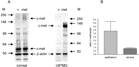
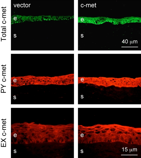
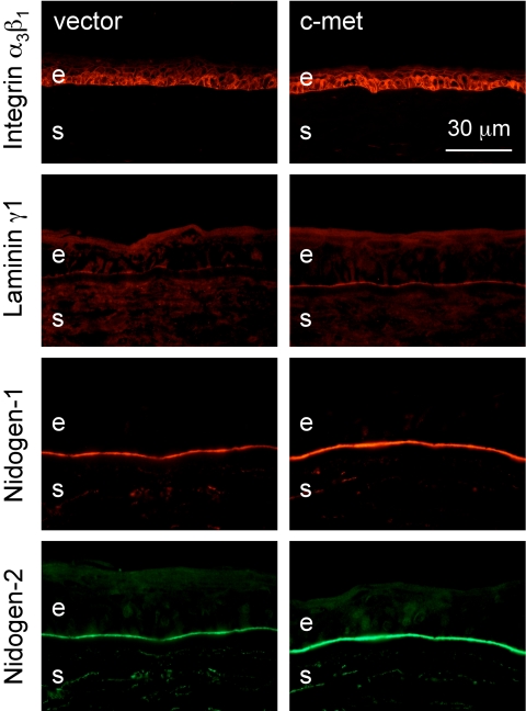
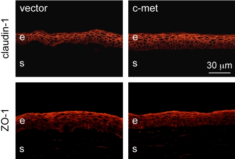
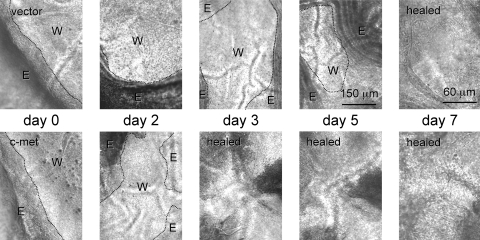
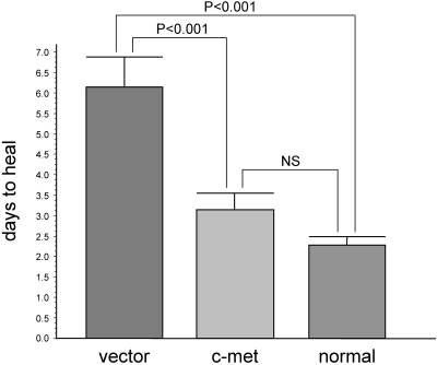
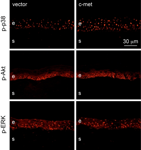
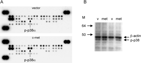
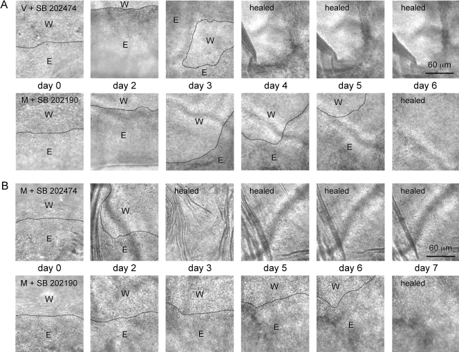
Purpose. Diabetic corneas display altered basement membrane and integrin markers, increased expression of proteinases, decreased hepatocyte growth factor (HGF) receptor, c-met proto-oncogene, and impaired wound healing. Recombinant adenovirus (rAV)-driven c-met overexpression in human organ-cultured corneas was tested for correction of diabetic abnormalities. Methods. Forty-six human corneas obtained postmortem from 23 donors with long-term diabetes (5 with diabetic retinopathy) were organ cultured and transduced with rAV-expressing c-met gene (rAV-cmet) under the cytomegalovirus promoter at approximately 10(8) plaque-forming units per cornea for 48 hours. Each control fellow cornea received control rAV (rAV expressing the beta-galactosidase gene or vector alone). After an additional 4 to 5 days of incubation, 5-mm epithelial wounds were created with n-heptanol, and healing was monitored. The corneas were analyzed afterward by immunohistochemistry and Western blot analysis. Signaling molecule expression and role was examined by immunostaining, phosphokinase antibody arrays, Western blot analysis, and inhibitor analysis. Results. rAV-cmet transduction led to increased epithelial staining for c-met (total, extracellular, and phosphorylated) and normalization of the patterns of select diabetic markers compared with rAV-vector-transduced control fellow corneas. Epithelial wound healing time in c-met-transduced diabetic corneas decreased twofold compared with rAV-vector-transduced corneas and became similar to normal. c-Met action apparently involved increased activation of p38 mitogen-activated protein kinase. c-Met transduction did not change tight junction protein patterns, suggesting unaltered epithelial barrier function. Conclusions. rAV-driven c-met transduction into diabetic corneas appears to restore HGF signaling, normalize diabetic marker patterns, and accelerate wound healing. c-Met gene therapy could be useful for correcting human diabetic corneal abnormalities.


Figures

Figure 1.
Figure 1.
rAV-cmet transduction into organ cultured human diabetic corneas and U87MG cells led to c-met overexpression. (A) Immunoblot analysis of total protein from corneas transduced with rAV-cmet and rAV-vector probed with rabbit pAb sc-161 (left), and from U87MG cell lysates (right). Arrows: 170-kDa full-length c-met, 140- to 145–kDa processed chain, and 55- to 60-kDa cell-associated fragment. Note that, in glioma cells, transduced c-met gets processed and mainly a 145-kDa chain is detected, whereas in the corneas, a full-length protein is present together with a prominent smaller band, apparently corresponding to a cell-associated kinase fragment. β-Actin was used to normalize protein loading. M, markers in kilodaltons. (B) Quantitative RT-PCR. On c-met gene transduction, its mRNA level increased approximately threefold in the epithelium, but stromal level did not change. Ratios to vector control are presented. β2-microglobulin was used as the housekeeping gene.
Figure 2.
Figure 2.
Transduction of c-met to organ cultured human diabetic corneas caused its increased expression and phosphorylation. Left: rAV-vector transduction; right: rAV-cmet transduction. In corneas transduced with rAV-cmet, there was increased epithelial staining for total c-met (revealed with a C-terminal rabbit pAb sc-10), tyrosine-phosphorylated (PY) c-met, and extracellular (EX) c-met. Indirect immunofluorescence of corneal sections. Sections of wounded and healed corneas are shown; the same exposure time was used for each pair of fellow corneas. e, epithelium; s, stroma.
Figure 3.
Figure 3.
c-Met transduction to organ cultured human diabetic corneas normalized expression patterns of select diabetic markers. Staining for integrin α3β1 became more homogeneous, and its intensity increased (top row). Staining for the BM component laminin-γ1 chain appeared after c-met transduction (second row). Staining for BM nidogen-1 and -2 (double labeling is shown in the two bottom rows) remained interrupted on vector transduction but became homogeneous after c-met transduction, similar to normal corneas. Indirect immunofluorescence of corneal sections. e, epithelium; s, stroma.
Figure 4.
Figure 4.
c-Met transduction did not change tight junction protein patterns. Top row: immunostaining for claudin-1 with typical membrane staining of all cell layers; bottom row: staining for ZO-1 mostly expressed in upper epithelial layers. None of these tight junction proteins appeared to change after c-met transduction. Indirect immunofluorescence of corneal sections. e, epithelium; s, stroma.
Figure 5.
Figure 5.
Dynamics of wound healing. A typical course of healing is presented for a c-met–transduced and vector-transduced cornea. rAV-vector–transduced diabetic cornea (top row) healed in 7 days, whereas rAV-met–transduced fellow cornea (bottom row) healed in 3 days. Pictures of live healing corneas are shown. Dashed line: shows the boundaries of the nonhealed wound region. W, wound zone; E, migrating epithelium.
Figure 6.
Figure 6.
c-Met overexpression led to a significant decrease in corneal epithelial wound healing time. The average healing time of rAV-vector–transduced corneas was 6.1 days, whereas rAV-cmet–transduced corneas healed in 3.1 days on average. For comparison, previous data on normal corneas are also shown. They healed in 2.3 days on average. Note that vector-treated diabetic corneas healed significantly slower than normal. c-Met transduction led to a significant decrease in healing time, bringing it close to normal. Statistical analysis of the time to complete healing (n = 7 for vector and c-met, and n = 13 for normal). Significance was determined using ANOVA with the Bonferroni post test.
Figure 7.
Figure 7.
c-Met overexpression leads to increased phosphorylation of p38 MAP kinase. Top row: increased epithelial staining for the phosphorylated form of p38 (p-p38). Staining for phosphorylated forms of Akt (p-Akt, middle row) or ERK (p-ERK, bottom row) kinases did not show noticeable change on c-met transduction. Indirect immunofluorescence of corneal sections. e, epithelium; s, stroma.
Figure 8.
Figure 8.
Phosphoproteomic array analysis of MAPKs. (A) On c-met transduction, phosphorylated p38α (p-p38α) was significantly (2.1-fold, n = 6, P < 0.05) increased compared with vector transduction. Array membranes were probed with total corneal protein, and signal was detected with chemiluminescence. (B) Validation of array results by Western blot analysis. Two separate cases are shown; in both of them p-p38 was increased on c-met transduction compared with vector alone. Mean increase in c-met versus vector was 1.7 ± 0.2 (n = 5, P < 0.04). Gel loading was normalized by β-actin. M, markers in kilodaltons.
Figure 9.
Figure 9.
Dynamics of wound healing on treatment with p38 inhibitor. (A, top row) rAV-vector–transduced diabetic cornea completely healed in 4 days in the presence of 10 μM of the inactive p38 inhibitor analogue SB 202474. Bottom row: rAV-met transduced fellow cornea healed in 6 days in the presence of 10 μM of the specific p38 inhibitor SB 202190. (B, top row) rAV-cmet–transduced diabetic cornea healed in 3 days in the presence of 10 μM SB 202474. Bottom row: rAV-met–transduced fellow cornea healed in 7 days in the presence of 10 μM SB 202190. Pictures of live corneas are shown. Analogue-treated corneas healed on average at 3.2 ± 0.4 days, compared with 5.7 ± 0.9 days for inhibitor-treated corneas (P < 0.05). p38 inhibitor thus abrogates c-met–induced acceleration of epithelial wound healing. Dashed line: shows the boundaries of the nonhealed wound region. W, wound zone; E, migrating epithelium.

Similar articles

Cited by

References

    1. Schultz RO, Van Horn DL, Peters MA, Klewin KM, Schutten WH. Diabetic keratopathy. Trans Am Ophthalmol Soc 1981;79:180–199 - PMC - PubMed
    1. Didenko TN, Smoliakova GP, Sorokin EL, Egorov VV. Clinical and pathogenetic features of neurotrophic corneal disorders in diabetes (in Russian). Vestn Oftalmol 1999;115:7–11 - PubMed
    1. Herse PR. A review of manifestations of diabetes mellitus in the anterior eye and cornea. Am J Optom Physiol Opt 1988;65:224–230 - PubMed
    1. Azar DT, Spurr-Michaud SJ, Tisdale AS, Gipson IK. Altered epithelial-basement membrane interactions in diabetic corneas. Arch Ophthalmol 1992;110:537–540 - PubMed
    1. Ljubimov AV, Huang Z, Huang GH, et al. Human corneal epithelial basement membrane and integrin alterations in diabetes and diabetic retinopathy. J Histochem Cytochem 1998;46:1033–1041 - PubMed

Publication types

MeSH terms

Substances