. 2010 Jan;38(3):750-63.
doi: 10.1093/nar/gkp1028. Epub 2009 Nov 19.

Adenovirus VA RNA-derived miRNAs target cellular genes involved in cell growth, gene expression and DNA repair

Affiliations

Adenovirus VA RNA-derived miRNAs target cellular genes involved in cell growth, gene expression and DNA repair

Oscar Aparicio et al. Nucleic Acids Res. 2010 Jan.

Abstract

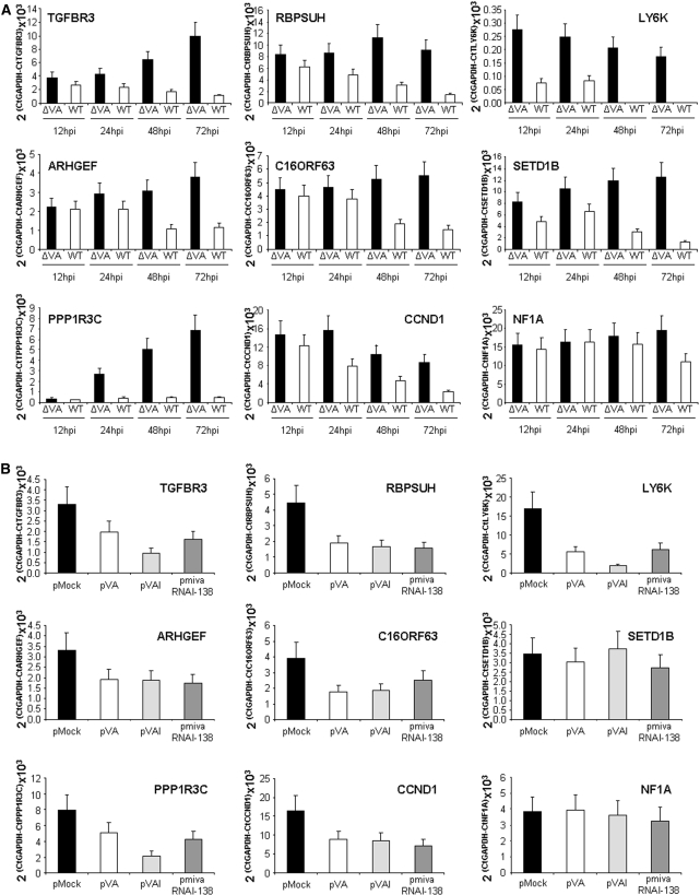
Adenovirus virus-associated (VA) RNAs are processed to functional viral miRNAs or mivaRNAs. mivaRNAs are important for virus production, suggesting that they may target cellular or viral genes that affect the virus cell cycle. To look for cellular targets of mivaRNAs, we first identified genes downregulated in the presence of VA RNAs by microarray analysis. These genes were then screened for mivaRNA target sites using several bioinformatic tools. The combination of microarray analysis and bioinformatics allowed us to select the splicing and translation regulator TIA-1 as a putative mivaRNA target. We show that TIA-1 is downregulated at mRNA and protein levels in infected cells expressing functional mivaRNAs and in transfected cells that express mivaRNAI-138, one of the most abundant adenoviral miRNAs. Also, reporter assays show that TIA-1 is downregulated directly by mivaRNAI-138. To determine whether mivaRNAs could target other cellular genes we analyzed 50 additional putative targets. Thirty of them were downregulated in infected or transfected cells expressing mivaRNAs. Some of these genes are important for cell growth, transcription, RNA metabolism and DNA repair. We believe that a mivaRNA-mediated fine tune of the expression of some of these genes could be important in adenovirus cell cycle.


Figures

Figure 1.
Figure 1.
(A) Work flow of the microarray analysis. RNA isolated from triplicates of HeLa cells mock transfected or transfected with pVA was hybridized to Affymetrix HG-U133-Plus2 microarrays containing 54 675 probes. Out of the 39 284 probes that showed expression, 4095 showed different expression between mock and pVA transfected cells. Expression differences were significant for 1384 probes which corresponded to 637 upregulated genes (730 probes) and 462 downregulated genes (654 probes) in the presence of VA RNAs. (B). Schematic of VAI RNA processing to viral miRNAs. The terminal nucleotide sequence of VA RNAI processed to viral miRNAs is indicated. The structure of the remaining VA RNAI is depicted with a black line. VA RNAI is processed to a minor 5′ miRNA and two major 3′ miRNAs (mivaRNAI-137 and -138). The cleavage site for each miRNA is indicated with an arrow.
Figure 2.
Figure 2.
Analysis of genes with putative mivaRNA target sequences whose expression is altered in the presence of VA RNAs. (A) Color code of the VA RNA microarray expression of the genes identified by TargetScan using VAI miRNAs as input sequences. Expression of the genes listed at the bottom of the table was detected with the indicated probes. The probes were spotted on microarrays hybridized with RNA isolated from mock transfected cells (pMock1 to 3) or cells transfected with pVA (pVA1-3), as indicated to the left. Expression levels range from red (higher expression) to green (lower expression), as gradiented to the right. The genes are upregulated (Up) or downregulated in pVA transfected cells, as indicated at the top of the figure. The genes shown are also identified by TargetScan with 5′mivaRNAI sequences (left) or 3′ mivaRNAI-138 sequences (right). (B) Summary of the analysis of 3′mivaRNA seed-complementary sequences in the genes with altered expression in the VA RNA microarray. The sequence, target seed-complementary sequence and relative abundance of major 3′mivaRNAs from VAI and VAII are shown. The table also indicates the number (n) and percentage (%) of genes with seed complementary sequences which are upregulated (up) or downregulated (down) in the presence of VA RNAs. (C) Bar graph showing the percentage of genes with altered expression in the pVA microarray and with seed complementary sequences. Seeds chosen are either total or complementary to positions 2–7 or 2–8 of the seed with or without an extra A. Seed target sequence from mivaRNAI-138, mivaRNAI-137 and mivaRNAII-138 is indicated at the top of the graphs. Plotted genes are either downregulated (down) or upregulated (up) in the presence of VA RNA.
Figure 3.
Figure 3.
Analysis of the putative miRNA target sequence in TIA-1 3′UTR. (A) mivaRNAI-138 target sequence in TIA-1 mRNA. Human (hs) TIA-1 3′UTR sequence close to the stop codon (bold) is shown. Non-conserved nucleotides of the same sequence in chimpanzee (pt) and mice (mm) are indicated. The putative binding showed between the mivaRNAI-138 and TIA-1 mRNA has been predicted by TargetScan. Base pairing is indicated with a line and GU wobbles are connected with a colon. The seed target is highlighted in bold and underlined. (B and C) Target sequences in TIA-1 3′UTR from cellular miRNAs. (B) TargetScan predicted binding between TIA-1 3′UTR and hsa-miR-892b, hsa-miR-339, hsa-miR-30c and hsa-miR-337-5p. (C) Seed complementary sequences to the miRNAs shown in (B) are boxed. mivaRNAI-138 seed binds to the seed target sequence of hsa-miR-30 and hsa-miR-337-5p.
Figure 4.
Figure 4.
TIA-1 mRNA is downregulated in the presence of VA RNA. (A and B) TIA-1 mRNA is downregulated in cells transfected with pVA. RNA was isolated from 293 (A) or HeLa (B) cells mock transfected or transfected with pVA, pVAI or pmivaRNAI-138. Then, GAPDH and TIA-1 mRNAs were quantified by RT–PCR and plotted for comparison. (C) TIA-1 mRNA is downregulated at late times postinfection with wild-type adenovirus. The 293 cells were infected with wild-type (AdWT) or a ΔVA mutant adenovirus and RNA was isolated at 6, 12, 24, 48 and 72 hpi. Quantitative RT–PCR was performed as indicated in (A). Error bars show standard deviations of three different experiments. Asterisk highlights significant differences while ns indicates non-significant differences.
Figure 5.
Figure 5.
TIA-1 protein is downregulated in the presence of VA RNA. (A) TIA-1 protein is downregulated in cells transfected with pVA. HeLa cells were transfected with control plasmids expressing a non-related gene (pMock) or a non-related shRNA (pshMock) or with pVA, pVAI or pmivaRNAI-138 plasmids. Extracts were collected at 48 h posttransfection and TIA-1 and U2AF65, used as a loading control, were evaluated by western blot analysis. (B) TIA-1 protein is downregulated in cells infected with wild-type adenovirus with functional 3′mivaRNAIs. 293 cells were transfected with antagomiRs that block 3′mivaRNAI function (2OmeAS3′) or with a mutant control (2OmetMut). These cells were infected with AdWT at 24 h posttransfection. Untransfected cells were also infected or mock infected, as a control. Extracts were collected at 24 hpi and TIA-1 and U2AF65 proteins were analyzed as in (A).
Figure 6.
Figure 6.
TIA-1 mRNA is a direct target of mivaRNAI-138. (A) Schematic of Renilla luciferase mRNA with TIA-1 mivaRNAI-138 target sequence (pRL-TIA-1) or with a matching seed complementary mutated sequence (pRL-TIA-1Mut). mivaRNAI-138 binding to the RNA is indicated for clarity. (B) mivaRNAI-138 decreases expression of a Renilla luciferase reporter mRNA with TIA-1 mivaRNAI-138 target sequences. HeLa cells were transfected with the plasmids expressing the Renilla constructs shown in (A), and a control plasmid or pVA, pVAI and pmivaRNAI-138 plasmids. A plasmid expressing a Firefly luciferase reporter was also co-transfected as a control. Luciferase activity was evaluated at 48 h posttransfection. Renilla luciferase expression in control cells was set to 100% and the percentage of Renilla expression in all cases was calculated accordingly. Error bars show standard deviations of three different experiments. Asterisk highlights significant differences while ns indicates non-significant differences.
Figure 7.
Figure 7.
mivaRNAs downregulate the expression of several genes. Expression of each gene was evaluated by quantitative RT–PCR in cells infected with wild-type or a ΔVA mutant adenovirus at 12, 24, 48 and 72 hpi (A) and in cells transfected with a control plasmid, pVA, pVAI or pmivaRNAI-138 (B). Error bars show standard deviations of three different measurements. Only representative genes are shown.

References

    1. Kim VN. MicroRNA biogenesis: coordinated cropping and dicing. Nat. Rev. Mol. Cell Biol. 2005;6:376–385. - PubMed
    1. Kim VN. Small RNAs just got bigger: Piwi-interacting RNAs (piRNAs) in mammalian testes. Genes Dev. 2006;20:1993–1997. - PubMed
    1. Eulalio A, Huntzinger E, Izaurralde E. Getting to the root of miRNA-mediated gene silencing. Cell. 2008;132:9–14. - PubMed
    1. Filipowicz W, Bhattacharyya SN, Sonenberg N. Mechanisms of post-transcriptional regulation by microRNAs: are the answers in sight? Nat. Rev. Genet. 2008;9:102–114. - PubMed
    1. Lingel A, Izaurralde E. RNAi: finding the elusive endonuclease. RNA. 2004;10:1675–1679. - PMC - PubMed

Publication types