. 2009 Dec 8;106(49):20800-5.
doi: 10.1073/pnas.0910550106. Epub 2009 Nov 20.

Mapping polypeptide interactions of the SecA ATPase during translocation

Affiliations

Mapping polypeptide interactions of the SecA ATPase during translocation

Benedikt W Bauer et al. Proc Natl Acad Sci U S A. .

Abstract

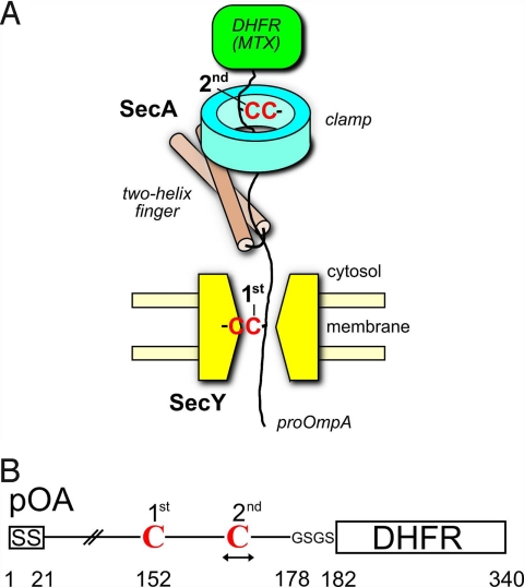
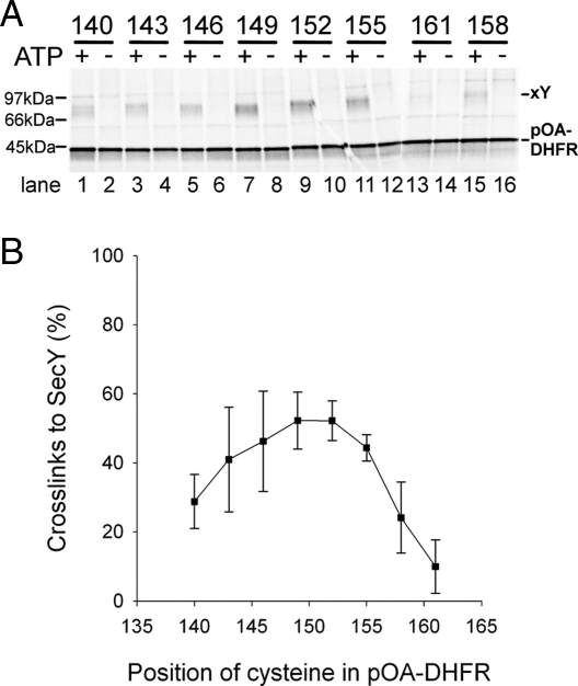
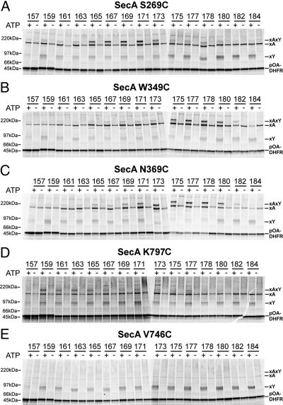
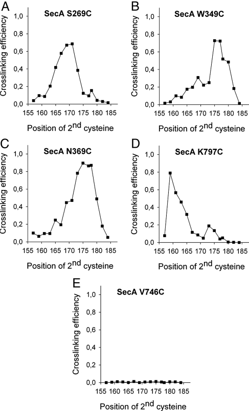
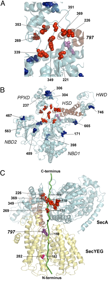
Many bacterial proteins, including most secretory proteins, are translocated across the plasma membrane by the interplay of the cytoplasmic SecA ATPase and a protein-conducting channel formed by the SecY complex. SecA catalyzes the sequential movement of polypeptide segments through the SecY channel. How SecA interacts with a broad range of polypeptide segments is unclear, but structural data raise the possibility that translocation substrates bind into a "clamp" of SecA. Here, we have used disulfide bridge cross-linking to test this hypothesis. To analyze polypeptide interactions of SecA during translocation, two cysteines were introduced into a translocation intermediate: one that cross-links to the SecY channel and the other one for cross-linking to a cysteine placed at various positions in SecA. Our results show that a translocating polypeptide is indeed captured inside SecA's clamp and moves in an extended conformation through the clamp into the SecY channel. These results define the polypeptide path during SecA-mediated protein translocation and suggest a mechanism by which ATP hydrolysis by SecA is used to move a polypeptide chain through the SecY channel.


Conflict of interest statement

The authors declare no conflict of interest.

Figures

Fig. 1.
Fig. 1.
Cross-linking strategy employing a translocation intermediate. (A) Scheme of the experimental strategy. Translocation of the C terminus of proOmpA was blocked by a fused dihydrofolate reductase (DHFR) domain that is folded in the presence of methotrexate (MTX). The fusion protein contains two cysteines (C), one for cross-linking to the SecY pore and a second at various positions for cross-linking to SecA mutants containing single cysteines (shown is a SecA with a cysteine in the clamp). (B) Scheme of the proOmpA (pOA)-DHFR construct. The position of the first cysteine (position 152) is kept constant whereas the position of the second is varied. GSGS is a linker sequence. SS, signal sequence.
Fig. 2.
Fig. 2.
Interaction of a translocation intermediate with the SecY pore. (A) pOA-DHFR containing a single cysteine at the indicated positions was synthesized in vitro in the presence of [35S]methionine. These substrates were incubated in the presence or absence of ATP with SecA lacking cysteines and proteoliposomes containing SecY with a single cysteine at position 282 in the pore ring. Disulfide bridge formation was induced with an oxidant. The samples were separated by SDS/PAGE and analyzed by autoradiography. Note that the samples 158 and 161 were loaded in the wrong order. The positions of free and SecY-cross-linked substrate (pOA-DHFR and xY) are indicated. (B) Quantification of three experiments performed as in A (mean and standard deviation). Shown is the percentage of substrate cross-linked to SecY [xY/(xY + pOA-DHFR)].
Fig. 3.
Fig. 3.
Probing interactions of a translocation intermediate with SecA. (A) Interaction of a substrate with the SecA clamp. pOA-DHFR containing a cysteine at positions 152 and a second cysteine at the indicated positions was synthesized in vitro in the presence of [35S]methionine. The substrate was incubated in the presence or absence of ATP with SecA containing a single cysteine in the clamp at position 269 and with proteoliposomes containing SecY with a cysteine at position 282. The samples were treated with an oxidant and analyzed by nonreducing SDS/PAGE and autoradiography. The positions of free, SecA-, SecY-, and double cross-linked substrate (pOA-DHFR, xA, xY, and xAxY) are indicated. (B) As in A but with a cysteine in the SecA clamp at position 349. (C) As in A but with a cysteine in the SecA clamp at position 369. (D) Interaction of a substrate with the two-helix finger of SecA. The experiments were performed as in A but with a cysteine at the SecA fingertip (position 797). (E) A cysteine randomly placed into SecA does not interact with the translocation intermediate. The experiments were performed as in A but with a cysteine at position 746 of SecA.
Fig. 4.
Fig. 4.
Quantification of the interactions of a translocation intermediate with SecA. (A–E) Quantified experiments from Fig. 3 A–E. The cross-linking efficiency is expressed as the ratio of double cross-links over the total SecY cross-links [xAxY/(xAxY + xY)].
Fig. 5.
Fig. 5.
Interaction sites with a translocating polypeptide mapped onto the T. maritima SecA structure (PDB accession 3DIN). (A) View from the cytosol of SecA positions that were tested for cross-linking to a translocating polypeptide chain. Cross-linking positions in the clamp and two-helix finger are shown as red and magenta balls, respectively. Positions that gave weak or no cross-links are shown in blue. The two-helix finger is shown in brown. (B) As in A but with a zoomed-out view. (C) Side view of the SecA-SecY structure with a modeled translocating pOA-DHFR substrate (pOA is shown in green; the DHFR domain was omitted for clarity). Positions in SecA's clamp and the two-helix finger, which could be cross-linked to the substrate, are shown as red and magenta balls, respectively. The cross-linking SecY pore residue is shown as pink balls. The star indicates an opening toward the cytosol.

References

    1. Rapoport TA. Protein translocation across the eukaryotic endoplasmic reticulum and bacterial plasma membranes. Nature. 2007;450:663–669. - PubMed
    1. van den Berg B, et al. X-ray structure of a protein-conducting channel. Nature. 2004;427:36–44. - PubMed
    1. Zimmer J, Nam Y, Rapoport TA. Structure of a complex of the ATPase SecA and the protein-translocation channel. Nature. 2008;455:936–943. - PMC - PubMed
    1. Tsukazaki T, et al. Conformational transition of Sec machinery inferred from bacterial SecYE structures. Nature. 2008;455:988–991. - PMC - PubMed
    1. Harris CR, Silhavy TJ. Mapping an interface of SecY (PrlA) and SecE (PrlG) by using synthetic phenotypes and in vivo cross-linking. J Bacteriol. 1999;181:3438–3444. - PMC - PubMed

Publication types

MeSH terms

LinkOut - more resources