. 2009 Dec 8;106(49):20960-5.
doi: 10.1073/pnas.0911405106. Epub 2009 Nov 20.

Activation of innate and humoral immunity in the peripheral nervous system of ALS transgenic mice

Affiliations

Activation of innate and humoral immunity in the peripheral nervous system of ALS transgenic mice

Isaac M Chiu et al. Proc Natl Acad Sci U S A. .

Abstract

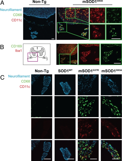
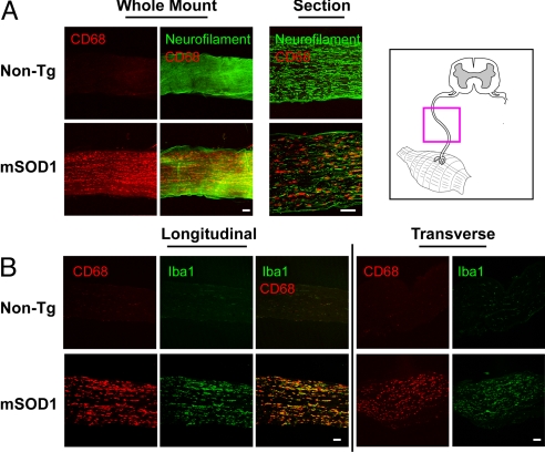
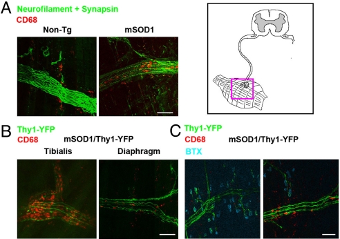
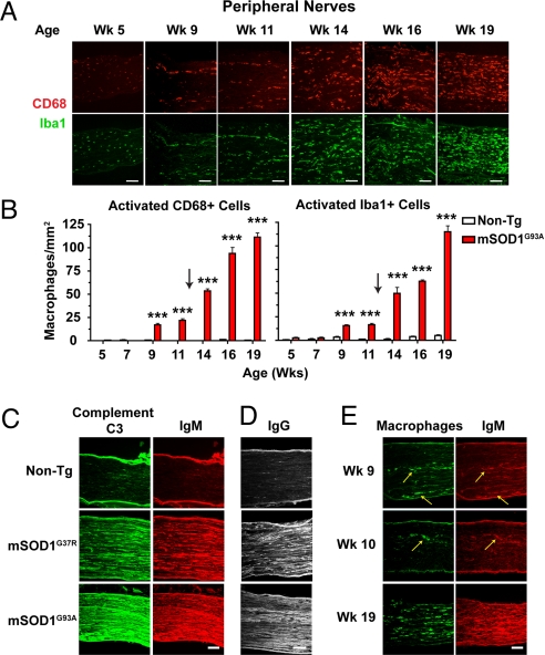
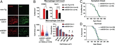
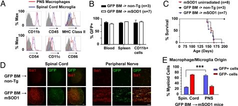
During injury to the nervous system, innate immune cells mediate phagocytosis of debris, cytokine production, and axon regeneration. In the neuro-degenerative disease amyotrophic lateral sclerosis (ALS), innate immune cells in the CNS are activated. However, the role of innate immunity in the peripheral nervous system (PNS) has not been well defined. In this study, we characterized robust activation of CD169/CD68/Iba1+ macrophages throughout the PNS in mutant SOD1(G93A) and SOD1(G37R) transgenic mouse models of ALS. Macrophage activation occurred pre-symptomatically, and expanded from focal arrays within nerve bundles to a tissue-wide distribution following symptom onset. We found a striking dichotomy for immune cells within the spinal cord and PNS. Flow cytometry and GFP bone marrow chimeras showed that spinal cord microglia were mainly tissue resident derived, dendritic-like cells, whereas in peripheral nerves, the majority of activated macrophages infiltrated from the circulation. Humoral antibodies and complement localized to PNS tissue in tandem with macrophage recruitment, and deficiency in complement C4 led to decreased macrophage activation. Therefore, cross-talk between nervous and immune systems occurs throughout the PNS during ALS disease progression. These data reveal a progressive innate and humoral immune response in peripheral nerves that is separate and distinct from spinal cord immune activation in ALS transgenic mice.


Conflict of interest statement

The authors declare no conflict of interest.

Figures

Fig. 1.
Fig. 1.
Morphologically activated macrophages expressing CD68, Iba1, CD11c, and CD169 accumulate between degenerating axons in ventral nerve roots of mutant SOD1 mice. (A) Both microglia in spinal cord and macrophages in ventral nerve roots of mSOD1G93A mice show significant expression of the myeloid activation marker, CD68 (green), and dendritic cell receptor, CD11c (red). Axons were labeled with anti-neurofilament (blue). Innate immune activation was absent in non-Tg sections. Magnified views (Insets) show images of representative microglia (Top) and nerve root macrophages (Bottom). (B) Anatomical schematic depicting spinal cord with ventral roots (Left). Magnification of mSOD1G93A section (Inset) shows nerve root macrophages expressing sialoadhesin, CD169 (green), and calcium adaptor, Iba1 (red). (C) Ventral roots in non-Tg, SOD1WT, and end-stage mSOD1G37R, mSOD1G93A mice stained for neurofilament (blue) and macrophage markers (CD11c, red; CD68, green). In mutant SOD1 mice, activated macrophages accumulate in spaces between degenerating axons. (Scale bars: 100 μm.)
Fig. 2.
Fig. 2.
Sciatic nerves in mutant SOD1 mice but not WT mice show intra-axonal activation of macrophages. (A) Whole-mount segments and longitudinal sections of distal sciatic nerve from end-stage mSOD1G93A and non-Tg litter-mates were stained for macrophages (CD68) and axons (neurofilament). For whole-mount stains, confocal microscopy images 100 μm into the nerve are shown as a composite. (B) Longitudinal and transverse sciatic nerve sections were stained for macrophage markers CD68 (red) and Iba1 (green). mSOD1G93A nerves at end stage show an abundance of activated, CD68/Iba1+ macrophages compared with non-Tg litter-mates. An anatomical schematic of peripheral nerves is shown (Upper Right). (Scale bars: 100 μm.)
Fig. 3.
Fig. 3.
Whole-mount muscle staining reveals macrophage infiltration within innervating axon fascicles. (A) Diaphragms from end-stage mSOD1G93A and non-Tg mice were stained for macrophages (CD68, red), axons (neurofilament, green), and synapses (synapsin, green). Intra-nerve macrophages were found in mSOD1G93A but not non-Tg muscles. (B) Muscles from end-stage mSOD1G93A/Thy1-YFP animals (YFP, green) were stained for macrophages (CD68, red). Activation of macrophages and degeneration of YFP axons was more extensive in the tibialis compared with diaphragm. (C) Triple staining for macrophages (CD68, red), axons (YFP, green), and motor endplates [bungarotoxin (BTX), blue]. Activated macrophages were found within mutant SOD1 axonal fascicles but not at neuromuscular synapses. (Scale bars: 100 μm.)
Fig. 4.
Fig. 4.
Age-dependent progression in PNS macrophage activation is accompanied by significant deposition of antibodies and complement. (A) Representative sciatic nerves from mSOD1G93A mice were stained for CD68/Iba1+ macrophages. At week 5, endoneural macrophages are morphologically un-activated. At weeks 9 and 11, macrophages accumulate in nerves, becoming activated morphologically, often assembling in rows. At weeks 14 to 19, activated macrophages spread throughout nerve parenchyma. (Scale bars: 100 μm.) (B) Quantification of progressive macrophage activation in sciatic nerves of mSOD1G93A and non-Tg mice (20× fields of non-consecutive 14 μm sections analyzed; mean ± SEM, ***, P < 0.001). (C) Week 19 mSOD1G93A, week 19 non-Tg, and 6-month-old mSOD1G37R sciatic nerve sections were stained for complement C3 (green) and antibody IgM (red). Strong deposition of complement and antibodies was observed in ALS Tg nerves. (D) Separate week-19 sections show anti-IgG reactivity in mSOD1G93A but not non-Tg nerves. (E) At presymptomatic weeks 9 and 10, antibody IgM (red) co-localizes in PNS tissue with strands of activated macrophages (CD68, green).
Fig. 5.
Fig. 5.
Complement C4 partially mediates macrophage activation during motor neuron degeneration. To ascertain the role of humoral immunity in ALS, mSOD1G93A mice were bred onto a complement C4-deficient background. (A) Representative images of Iba1/CD68+ macrophages in end-stage mSOD1C4+/+ and mSOD1C4−/− sciatic nerves, with age-matched non-Tg nerves. (Scale bars: 100 μm.) (B) Quantification analysis of CD68+ and Iba1+ macrophages in end-stage mSOD1C4+/+ (n = 5), mSOD1C4−/− sciatic nerves (n = 5). Individual macrophage size data (CD68+) are shown in vertical scatter plot and histogram analysis (mean ± SEM, *, P < 0.05 by t test). (C and D) Kaplan Meier curves of symptom onset and survival in mSOD1C4+/+ (n = 25; male, n = 14; female, n = 11) and mSOD1C4−/− mice (n = 35; male, n = 18; female, n = 17).
Fig. 6.
Fig. 6.
Flow cytometry and GFP chimeras demonstrate unique nature and origin for ALS PNS macrophages compared with spinal cord microglia. (A) Sciatic nerve macrophages and spinal cord microglia from end-stage mSOD1G93A mice were compared by FACS for MHC class II, CD54 (ICAM-1), CD11c, and CD86. Histograms show surface expression of PNS macrophages (red) and spinal cord microglia (blue) relative to isotype controls for PNS macrophages (gray). (B-E) mSOD1G93A (n = 8) or non-Tg (n = 3) mice were transplanted at 7 weeks with BM from EGFP mice (GFP BM). Un-irradiated mSOD1G93A litter-mates (n = 7) were analyzed in parallel. (B) FACS analysis of GFP chimerism for blood leukocytes and total and CD11b+ splenocytes in transplanted mice at end-stage. (C) Kaplan–Meier survival analysis of BM-transplanted and un-irradiated mice. (D) In spinal cord, Iba1+ microglia are mainly negative for GFP, indicating tissue resident origin. In contrast, sciatic nerve sections imaged for Iba1+ macrophages show significant co-expression of GFP, indicating a BM origin. (Scale bars: 100 μm.) (E) Quantification of BM-derived spinal cord microglia and PNS macrophages in mutant SOD1/GFP mice (***, P < 0.001; n = 4).

References

    1. Rosen DR, et al. Mutations in Cu/Zn superoxide dismutase gene are associated with familial amyotrophic lateral sclerosis. Nature. 1993;362:59–62. - PubMed
    1. Gurney ME, et al. Motor neuron degeneration in mice that express a human Cu,Zn superoxide dismutase mutation. Science. 1994;264:1772–1775. - PubMed
    1. Bruijn LI, et al. ALS-linked SOD1 mutant G85R mediates damage to astrocytes and promotes rapidly progressive disease with SOD1-containing inclusions. Neuron. 1997;18:327–338. - PubMed
    1. Mcgeer P, Mcgeer E. Inflammatory processes in amyotrophic lateral sclerosis. Muscle Nerve. 2002;26:459–470. - PubMed
    1. Hall ED, Oostveen JA, Gurney ME. Relationship of microglial and astrocytic activation to disease onset and progression in a transgenic model of familial ALS. Glia. 1998;23:249–256. - PubMed

Publication types

MeSH terms