Stem cell signaling in Arabidopsis requires CRN to localize CLV2 to the plasma membrane
- PMID: 19933383
- PMCID: PMC2799354
- DOI: 10.1104/pp.109.149930
Stem cell signaling in Arabidopsis requires CRN to localize CLV2 to the plasma membrane
Abstract
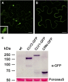
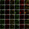
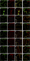
Stem cell number in shoot and floral meristems of Arabidopsis (Arabidopsis thaliana) is regulated by the CLAVATA3 (CLV3) signaling pathway. Perception of the CLV3 peptide requires the receptor kinase CLV1, the receptor-like protein CLV2, and the kinase CORYNE (CRN). Genetic analysis suggested that CLV2 and CRN act together and in parallel with CLV1. We studied the intracellular localization of receptor fusions with fluorescent protein tags and their capacities for interaction via efficiency of fluorescence resonance energy transfer. We found that CLV2 and CRN require each other for export from the endoplasmic reticulum and localization to the plasma membrane (PM). CRN readily forms homomers and interacts with CLV2 through the transmembrane domain and adjacent juxtamembrane sequences. CLV1 forms homomers independently of CLV2 and CRN at the PM. We propose that the CLV3 signal is perceived by a tetrameric CLV2/CRN complex and a CLV1 homodimer that localize to the PM and can interact via CRN.
Figures


Similar articles
-
Analysis of interactions among the CLAVATA3 receptors reveals a direct interaction between CLAVATA2 and CORYNE in Arabidopsis.Plant J. 2010 Jan;61(2):223-33. doi: 10.1111/j.1365-313X.2009.04049.x. Epub 2009 Oct 16. Plant J. 2010. PMID: 19843317
-
The receptor kinase CORYNE of Arabidopsis transmits the stem cell-limiting signal CLAVATA3 independently of CLAVATA1.Plant Cell. 2008 Apr;20(4):934-46. doi: 10.1105/tpc.107.057547. Epub 2008 Apr 1. Plant Cell. 2008. PMID: 18381924 Free PMC article.
-
CLAVATA2 forms a distinct CLE-binding receptor complex regulating Arabidopsis stem cell specification.Plant J. 2010 Sep;63(6):889-900. doi: 10.1111/j.1365-313X.2010.04295.x. Plant J. 2010. PMID: 20626648 Free PMC article.
-
CLAVATA signaling pathway receptors of Arabidopsis regulate cell proliferation in fruit organ formation as well as in meristems.Genetics. 2011 Sep;189(1):177-94. doi: 10.1534/genetics.111.130930. Epub 2011 Jul 29. Genetics. 2011. PMID: 21705761 Free PMC article.
-
CLAVATA3, a plant peptide controlling stem cell fate in the meristem.Peptides. 2021 Aug;142:170579. doi: 10.1016/j.peptides.2021.170579. Epub 2021 May 24. Peptides. 2021. PMID: 34033873 Review.
Cited by
-
GIGANTEA recruits the UBP12 and UBP13 deubiquitylases to regulate accumulation of the ZTL photoreceptor complex.Nat Commun. 2019 Aug 21;10(1):3750. doi: 10.1038/s41467-019-11769-7. Nat Commun. 2019. PMID: 31434902 Free PMC article.
-
Receptor-like protein kinases in plant reproduction: Current understanding and future perspectives.Plant Commun. 2021 Dec 29;3(1):100273. doi: 10.1016/j.xplc.2021.100273. eCollection 2022 Jan 10. Plant Commun. 2021. PMID: 35059634 Free PMC article. Review.
-
HANABA TARANU regulates the shoot apical meristem and leaf development in cucumber (Cucumis sativus L.).J Exp Bot. 2015 Dec;66(22):7075-87. doi: 10.1093/jxb/erv409. Epub 2015 Aug 28. J Exp Bot. 2015. PMID: 26320238 Free PMC article.
-
Identification and characterization of the Populus trichocarpa CLE family.BMC Genomics. 2016 Mar 2;17:174. doi: 10.1186/s12864-016-2504-x. BMC Genomics. 2016. PMID: 26935217 Free PMC article.
-
CLAVATA Signaling Ensures Reproductive Development in Plants across Thermal Environments.Curr Biol. 2021 Jan 11;31(1):220-227.e5. doi: 10.1016/j.cub.2020.10.008. Epub 2020 Nov 5. Curr Biol. 2021. PMID: 33157018 Free PMC article.
References
-
- Albertazzi L, Arosio D, Marchetti L, Ricci F, Beltram F (2009) Quantitative FRET analysis with the EGFP-mCherry fluorescent protein pair. Photochem Photobiol 85 287–297 - PubMed
-
- Ali GS, Prasad KV, Day I, Reddy AS (2007) Ligand-dependent reduction in the membrane mobility of FLAGELLIN SENSITIVE2, an Arabidopsis receptor-like kinase. Plant Cell Physiol 48 1601–1611 - PubMed
-
- Boller T, Felix G (2009) A renaissance of elicitors: perception of microbe-associated molecular patterns and danger signals by pattern-recognition receptors. Annu Rev Plant Biol 60 379–406 - PubMed
-
- Brand U, Fletcher JC, Hobe M, Meyerowitz EM, Simon R (2000) Dependence of stem cell fate in Arabidopsis on a feedback loop regulated by CLV3 activity. Science 289 617–619 - PubMed
-
- Chan FK, Chun HJ, Zheng L, Siegel RM, Bui KL, Lenardo MJ (2000) A domain in TNF receptors that mediates ligand-independent receptor assembly and signaling. Science 288 2351–2354 - PubMed
Publication types
MeSH terms
Substances
LinkOut - more resources
Full Text Sources
Other Literature Sources
Medical
Molecular Biology Databases
Miscellaneous

