

Imaging findings in a fatal case of pandemic swine-origin influenza A (H1N1)
- PMID: 19933640
- PMCID: PMC2788497
- DOI: 10.2214/AJR.09.3365
Imaging findings in a fatal case of pandemic swine-origin influenza A (H1N1)
Abstract
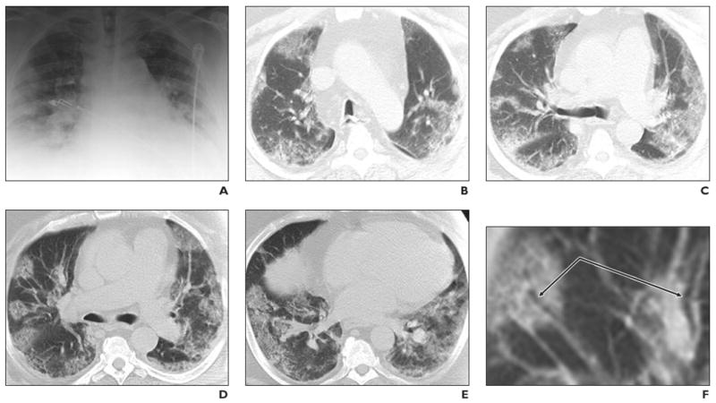
Objective: Although most cases of swine-origin influenza A (H1N1) virus (S-OIV) have been self-limited, fatal cases raise questions about virulence and radiology's role in early detection. We describe the radiographic and CT findings in a fatal S-OIV infection.
Conclusion: Radiography showed peripheral lung opacities. CT revealed peripheral ground-glass opacities suggesting peribronchial injury. These imaging findings raised suspicion of S-OIV despite negative H1N1 influenza rapid antigen test results from two nasopharyngeal swabs; subsequently, those results were proven to be false-negatives by reverse transcriptase polymerase chain reaction. This case suggests a role for CT in the early recognition of severe S-OIV.
Figures
Comment in
-
Conventional wisdom: unconventional virus.AJR Am J Roentgenol. 2009 Dec;193(6):1486-7. doi: 10.2214/AJR.09.3758. AJR Am J Roentgenol. 2009. PMID: 19933637 No abstract available.
References
-
- Perez-Padilla R, de la Rosa-Zamboni D, Ponce de Leon S, et al. INER Working Group on Influenza. Pneumonia and respiratory failure from swine-origin influenza A (H1N1) in Mexico. N Engl J Med. 2009;361:680–689. - PubMed
-
- Qureshi NR, Hien TT, Farrar J, Gleeson FV. The radiologic manifestations of H5N1 avian influenza. J Thorac Imaging. 2006;21:259–264. - PubMed
-
- Ebell MH, White LL, Casault T. A systematic review of the history and physical examination to diagnose influenza. J Am Board Fam Pract. 2004;17:1–5. - PubMed
-
- Centers for Disease Control and Prevention (CDC) Update: novel influenza A (H1N1) virus infections—worldwide, May 6, 2009. MMWR Morb Mortal Wkly Rep. 2009;58:453–458. - PubMed
Publication types
MeSH terms
Grants and funding
LinkOut - more resources
Full Text Sources
Other Literature Sources
Medical
