. 2010 Jan 1;588(Pt 1):117-24.
doi: 10.1113/jphysiol.2009.181511. Epub 2009 Nov 23.

Quantitative characterization of low-threshold mechanoreceptor inputs to lamina I spinoparabrachial neurons in the rat

Affiliations

Quantitative characterization of low-threshold mechanoreceptor inputs to lamina I spinoparabrachial neurons in the rat

David Andrew. J Physiol. .

Abstract

It has been suggested that primary afferent C-fibres that respond to innocuous tactile stimuli are important in the sensation of pleasurable touch. Although it is known that C-tactile fibres terminate in the substantia gelatinosa (lamina II) of the spinal cord, virtually all of the neurons in this region are interneurons, and currently it is not known how impulses in C-mechanoreceptors are transmitted to higher centres. In the current study, I have tested the quantitative response properties of 'wide dynamic range' projection neurons in lamina I of the spinal cord to graded velocity brushing stimuli to identify whether low-threshold mechanoreceptor input to these neurons arises from myelinated or umyelinated nerve fibres. Graded velocity brushing stimuli (6.6-126 cm s(-1)) were used to characterize the mechanoreceptor inputs to 'wide dynamic range' neurons in lamina I of the dorsal horn that had axons that projected to the contralateral parabrachial nucleus. The most effective tactile stimuli for activation of 'wide dynamic range' lamina I spinoparabrachial neurons were low velocity brush strokes: peak discharge occurred at a mean velocity of 9.2 cm s(-1) (range 6.6-20.4 cm s(-1), s.d. 5.0 cm s(-1)), and declined exponentially as brush velocity increased. The data indicate that C-fibres, but not A-fibres, conveyed low-threshold mechanoreceptor inputs to lamina I projection neurons.


Figures
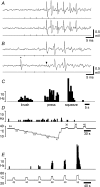
Figure 1
Figure 1. Identification of ‘wide dynamic range’ lamina I spinoparabrachial neurons in vivo
A, pair of traces showing 1-for-1 following of a train of 6 antidromic electrical stimuli (40 μA, 1 ms, 250 Hz; vertical ticks) delivered from a stimulating electrode in the contralateral parabrachial nucleus. The conduction distance was 87 mm. B, collision of the first antidromic impulse in a train of 3 (150 Hz, upper trace) when an orthodromic impulse (asterisk, lower trace) occurred within the critical interval. The arrowhead indicates the point at which the first antidromic response should have occurred. Vertical ticks indicate the timing of the antidromic stimuli. C, peri-stimulus time histogram showing the response of the neuron shown above to innocuous (brushing with a hand-held brush; velocity ∼1 cm s−1) and noxious mechanical stimuli. D, histogram showing the response of the same neuron to graded cooling stimuli, applied with a feedback-controlled Peltier element. E, response of the same unit to graded heat.
Figure 3
Figure 3. Quantitative velocity encoding by ‘wide dynamic range’ lamina I spinoparabrachial neurons
A, simulus–response curves of velocity encoding for the individual neurons, as well as the population mean (•, thick line). The population response was well fitted by a first-order exponential decay function of the form y= 48.7e−0.06x (r2= 0.93). B, the same data as A, but transformed logarithmically. The population response was well fitted by a straight line function of the form y=−1.87x+ 3.33 (r2= 0.93). C, stimulus–response curves of the velocity dependence of the mean number of action potentials evoked per stimulus, as well as the population mean (•, thick line). As can be seen, higher velocity stimuli evoked fewer action potentials, due to shorter stimulus duration. D, stimulus–response curves of response probability as a function of velocity for the individual neurons, as well as the population mean (•, thick line). For each neuron at each velocity the proportion of stimuli that evoked any action potentials was determined, and the failure probability, i.e. the proportion of trials that evoked no response calculated. As can be seen, as velocity increased, the probability of response failure increased.
Figure 2
Figure 2. Responses to brushing stimuli
A, raw responses from a typical ‘wide dynamic range’ lamina I spinoparabrachial neuron to repeated brushing at the lowest velocity tested (6.6 cm s−1). The first, then every other response to a series of 10 stimulus repetitions are shown. A marker trace (bottom) from a photocell indicates stimulus timing. As can be seen, there is a gradual reduction in response as the stimulus is repeated, similar to primary afferent C-fibre mechanoreceptors. B, responses of the same cell to 4 other of the 10 different brush velocities that were tested. Responses to the 1st, 5th and 10th stimuli are shown, with each action potential represented by a vertical tick mark. As can be seen, increasing stimulus velocity caused a progressive reduction in response.
Figure 4
Figure 4. Differentiation of the low-threshold inputs to lamina I neurons from low-threshold inputs to lamina III neurons
A, raw responses from a single lamina III spinoparabrachial neuron showing collision of the first antidromic impulse in a train of 3 (150 Hz, upper trace) when an orthodromic impulse (asterisk, lower trace) occurred within the critical interval. The arrowhead indicates the point at which the first antidromic response should have occurred. B, stimulus–response curves from the population of lamina I neurons and the single ‘wide dynamic range’ lamina III neuron to graded velocity brushing. Note how evoked discharge increases for the lamina III neuron. C, same data as in B but transformed logarithmically.

Comment in

  • Why a soft touch can hurt.
    Craig AD. Craig AD. J Physiol. 2010 Jan 1;588(Pt 1):13. doi: 10.1113/jphysiol.2009.185116. J Physiol. 2010. PMID: 20045900 Free PMC article. No abstract available.

Similar articles

Cited by

References

    1. Alden M, Besson JM, Bernard JF. Organization of the efferent projections from the pontine parabrachial area to the bed nucleus of the stria terminalis and neighboring regions: a PHA-L study in the rat. J Comp Neurol. 1994;341:289–314. - PubMed
    1. Al-Khater KM, Todd AJ. Collateral projections of neurons in laminae I, III and IV of rat spinal cord to thalamus, periaqueductal grey matter, and lateral parabrachial area. J Comp Neurol. 2009;515:629–646. - PMC - PubMed
    1. Andrew D. Sensitization of lamina I spinoparabrachial neurons parallels heat hyperalgesia in the chronic constriction injury model of neuropathic pain. J Physiol. 2009;587:2005–2018. - PMC - PubMed
    1. Bernard JF, Alden M, Besson JM. The organization of the efferent projections from the pontine parabrachial area to the amygdaloid complex: a Phaseolus vulgaris leucoagglutinin (PHA-L) study in the rat. J Comp Neurol. 1993;329:201–229. - PubMed
    1. Bessou P, Burgess PR, Perl ER, Taylor CB. Dynamic properties of mechanoreceptors with unmyelinated (C) fibres. J Neurophysiol. 1971;34:116–131. - PubMed

Publication types