. 2010 Feb;78(2):639-50.
doi: 10.1128/IAI.01043-09. Epub 2009 Nov 23.

Transitions in oral and intestinal microflora composition and innate immune receptor-dependent stimulation during mouse development

Affiliations

Transitions in oral and intestinal microflora composition and innate immune receptor-dependent stimulation during mouse development

Mizuho Hasegawa et al. Infect Immun. 2010 Feb.

Abstract

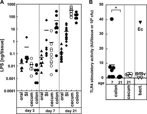
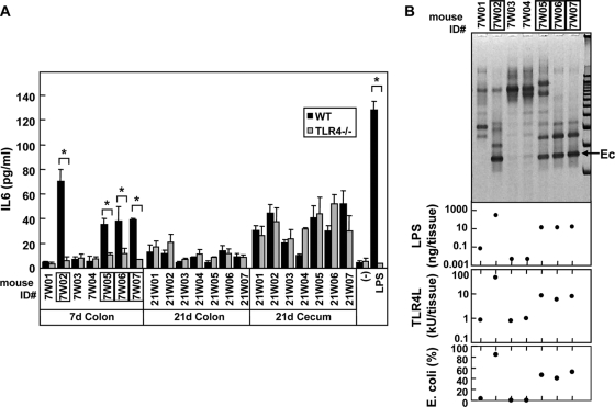
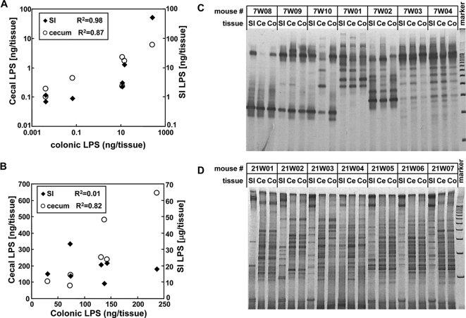
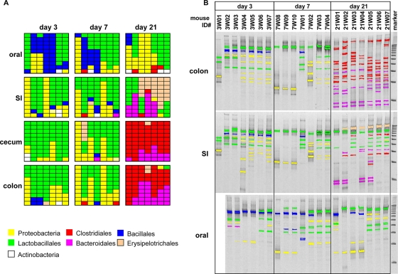
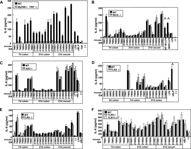
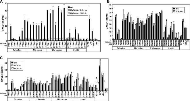
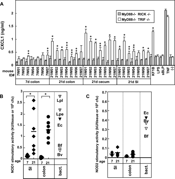
Commensal bacteria possess immunostimulatory activities that can modulate host responses to affect development and homeostasis in the intestine. However, how different populations of resident bacteria stimulate the immune system remains largely unknown. We characterized here the ability of intestinal and oral microflora to stimulate individual pattern recognition receptors (PRRs) in bone marrow-derived macrophages and mesothelial cells. The intestinal but not oral microflora elicited age- and cell type-specific immunostimulation. The immunostimulatory activity of the intestinal microflora varied among individual mice but was largely mediated via Toll-like receptor 4 (TLR4) during breast-feeding, whereas it became TLR4 independent after weaning. This transition was associated with a change from a microflora rich in TLR4-stimulatory proteobacteria to one dominated by Bacteroidales and/or Clostridiales that poorly stimulate TLR4. The major stimulatory activity of the intestinal microflora was still intact in NOD1-, NOD2-, TLR2-, TLR4-, TLR5-, TLR9-, TLR11-, ASC-, or RICK-deficient cells but still relied on the adaptor MyD88. These studies demonstrate a transition in the intestinal microflora accompanied by a dynamic change of its ability to stimulate different PRRs which control intestinal homeostasis.


Figures

FIG. 1.
FIG. 1.
Stimulation of BMM by oral and intestinal microflora. BMM were stimulated with tissue homogenates derived from the oral cavity, small intestine (SI), and colon from different 3-, 7-, and 21-day-old mice (n = 7) or adult germfree (gf) mice. The amounts of IL-6 (A), CCL5 (B), and IL-10 (C) in culture supernatants were determined by enzyme-linked immunosorbent assay (ELISA). (D) Correlation between IL-6 and CCL5 (•) levels and between IL-6 and IL-10 levels (○). (E) Correlation between total bacterial number and levels of CCL5 (•) and IL-10 (○). (F) Secretion of CXCL1 by mesothelial cells stimulated with tissue homogenates from the indicated sites from 3-, 7-, and 21-day-old B6 mice. The amounts of CXCL1 in culture supernatants were determined by ELISA. Each symbol represents mean value of triplicate cultures from individual mice. The standard deviations (SD) of triplicate cultures for individual mice averaged less than 19%. Bars denote mean values. *, P < 0.001.
FIG. 2.
FIG. 2.
Difference between the LPS content and TLR4-immunostimulatory activity in oral and intestinal tissues. (A) Total amounts of LPS on day 3, 7, and 21 in oral tissue (▴), small intestine (SI; ⧫), cecum (○), and colon (•) were determined by LAL assay. (B) TLR4-stimulatory activity in oral and intestinal tissues. The levels of TLR4-stimulatory activity in cecal and colonic homogenates (n = 7) at days 7 and 21 were determined by the bioassay using HEK293T cells expressing TLR4 and MD-2. Bars denote mean values. As reference controls, the LPS levels of 109 CFU of Gram-negative bacteria, E. coli (Ec), B. fragilis (Bf), and B. vulgatus (Bv) are shown. *, P < 0.001.
FIG. 3.
FIG. 3.
TLR4-dependent macrophage-stimulatory activity in 7-day-old mice is associated with E. coli predominance. (A) BMM from WT and TLR4−/− mice were treated with the medium alone (−) and heat-inactivated homogenates (diluted 100-fold) from the indicated tissues of 3-, 7-, and 21-day-old B6 mice. Stimulation with 100 ng of LPS/ml is also shown as a control. The amounts of IL-6 secreted by BMM after 12 h of stimulation were determined by ELISA. The data are represented as means ± the SD. *, P < 0.001. (B) Comparison of dominant bacteria LPS content and TLR4-stimulatory activity in the small intestinal microflora on day 7. The major bacterial species from individual mice were analyzed by DGGE (upper panel) and compared to the LPS content and TLR4-stimulatory activity (second and third panels). Ec, band corresponding to E. coli as determined by sequencing. The percentage of E. coli in the whole bacterial population of the colon(bottom panel) was determined by real-time PCR as described in Materials and Methods. The numbers of mice that possessed E. coli dominant microflora are boxed.
FIG. 4.
FIG. 4.
Transition of dominant bacteria in intestinal microflora during mouse development. (A and B) Correlation between LPS levels within the small intestine (SI), cecum (Ce), and colon (Co) on day 7 (A) and day 21 (B). (C and D) Comparison of dominant bacterial species in the intestine by DGGE analysis on day 7 (C) and day 21 (D). All mouse sets used in panels A and B were the same as those used in panels C and D, respectively.
FIG. 5.
FIG. 5.
Identification of dominant bacteria in oral and intestinal microflora during mouse development. (A) The bacterial DNA prepared from homogenates of oral and intestinal tissues were amplified with 16S rRNA gene consensus primer set. A total of 876 clones were randomly isolated and sequenced from 16S rRNA gene libraries generated from each microflora. The identity and homology of each clone were determined by using the BLASTN program. First, 10 clones from each sample are shown by the distinct colors corresponding to the taxonomic groups. The full description of bacteria species are shown in Table S1 in the supplemental material. (B) The DNA was further amplified with the GC-clump-linked consensus primer and separated by DGGE with DNA 100-bp ladder markers. The major DNA fragments were recovered from the gel and sequenced as described in Materials and Methods. The identities of bacteria were determined by homology search using the BLASTN program. The DNA fragments of characterized bacteria are depicted in the same colors as in panel A. The original images and full descriptions of the bacterial species are shown in Fig. S2 and Table S2, respectively, in the supplemental material.
FIG. 6.
FIG. 6.
Dominant role of MyD88/TRIF signaling in immune responses of BMM to intestinal microflora. BMM from a control WT, MyD88−/− TRIF−/− (A), RICK−/− (B), ASC−/− (C), TLR2−/− (D), TLR9−/− (E), TLR5−/−, or TLR11−/− (F) mice were stimulated with medium alone (−) or heat-inactivated homogenates from the indicated tissues of 7- and 21-day-old B6 mice. Stimulation with 10 ng of LPS/ml, 5 μg of KF1B/ml plus 10 ng of LPS/ml, 5 μg of MDP/ml plus 10 ng of LPS/ml, or 1 μg of sBLP/ml is shown as a control. The amounts of IL-6 in culture supernatants after 12 h of stimulation were determined by ELISA. The data are represented as means ± the SD. * and **, P < 0.001 and P < 0.01, respectively.
FIG. 7.
FIG. 7.
Dominant role of MyD88 in immune responses of mesothelial cells to intestinal microflora. Mesothelial cells from WT, MyD88−/− TRIF−/−, MyD88−/− RICK−/− (A), TLR9−/− (B), and RICK−/− and NOD1−/− (C) mice were treated with medium alone (−) or heat-inactivated homogenates (diluted 100-fold) from the indicated tissues of 7- and 21-day-old B6 mice. Stimulation with 100 ng of LPS/ml, 1 μg of sBLP/ml, or 10 ng of TNF-α/ml is shown as a control. The amounts of CXCL1 in culture supernatants after 12 h of stimulation were determined by ELISA. The data are represented as mean ± the SD. * and **, P < 0.001 and P < 0.01, respectively, compared to WT and mutant cells.
FIG. 8.
FIG. 8.
Secondary contribution of RICK and NOD1 in immune responses of mesothelial cells to intestinal microflora. (A) Mesothelial cells from MyD88−/−TRIF−/− and MyD88−/− RICK−/− mice were treated with medium alone (−) or heat-inactivated homogenates (diluted 100-fold) from indicated tissue of 7- and 21-day-old B6 mice. Stimulation with 100 ng of LPS/ml, 1 μg of sBLP/ml, 5 μg of KF1B/ml, 10 ng of tumor necrosis factor alpha (TNF-α)/ml is shown as a control. The amounts of CXCL1 in culture supernatants after 12 h of stimulation were determined by ELISA. The results were derived from the same experiment shown in Fig. 7B but on a different scale to reveal differences in CXCL1 secretion between MyD88−/− TRIF−/− and MyD88−/− RICK−/− mesothelial cells. The data are represented as means ± the SD. (B and C) Levels of NOD1 and NOD2 stimulatory activities in oral and intestinal tissues. The levels of NOD1 (B) and NOD2 (C) stimulatory activity in cecal and colonic homogenates (n = 7) at days 7 and 21 were determined by a bioassay using HEK293T cells expressing NOD1 or NOD2. The NOD1 stimulatory levels of 109 CFU of the mesoDAP-type peptidoglycan-containing bacteria Lactobacillus plantarum (Lpl), L. pentosus (Lpe), E. coli (Ec), B. fragilis (Bf), and B. vulgatus (Bv) or the NOD2 stimulatory levels of 109 CFU of E. coli (Ec), B. fragilis (Bf), and B. vulgatus (Bv) are shown as controls.

References

    1. Reference deleted.
    1. Artis, D. 2008. Epithelial-cell recognition of commensal bacteria and maintenance of immune homeostasis in the gut. Nat. Rev. Immunol. 8:411-420. - PubMed
    1. Atarashi, K., J. Nishimura, T. Shima, Y. Umesaki, M. Yamamoto, M. Onoue, H. Yagita, N. Ishii, R. Evans, K. Honda, and K. Takeda. 2008. ATP drives lamina propriaT(H)17 cell differentiation. Nature 455:808-812. - PubMed
    1. Bartosch, S., A. Fite, G. T. Macfarlane, and M. E. McMurdo. 2004. Characterization of bacterial communities in feces from healthy elderly volunteers and hospitalized elderly patients by using real-time PCR and effects of antibiotic treatment on the fecal microbiota. Appl. Environ. Microbiol. 70:3575-3581. - PMC - PubMed
    1. Bouskra, D., C. Brézillon, M. Bérard, C. Werts, R. Varona, I. G. Boneca, and G. Eberl. 2008. Lymphoid tissue genesis induced by commensals through NOD1 regulates intestinal homeostasis. Nature 456:507-510. - PubMed

Publication types

Substances