. 2010 Feb;78(2):629-38.
doi: 10.1128/IAI.01019-09. Epub 2009 Nov 23.

Protease-activated receptor 2 has pivotal roles in cellular mechanisms involved in experimental periodontitis

Affiliations

Protease-activated receptor 2 has pivotal roles in cellular mechanisms involved in experimental periodontitis

David M Wong et al. Infect Immun. 2010 Feb.

Abstract

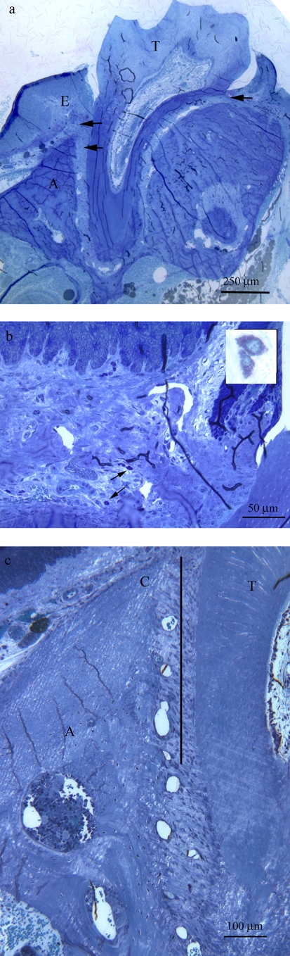
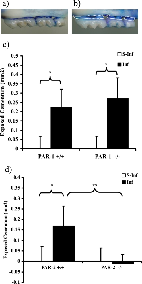
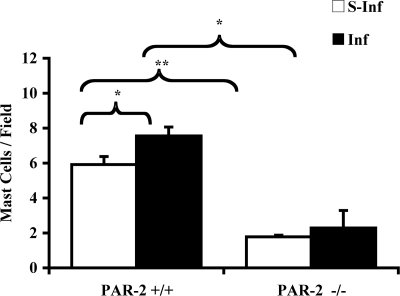
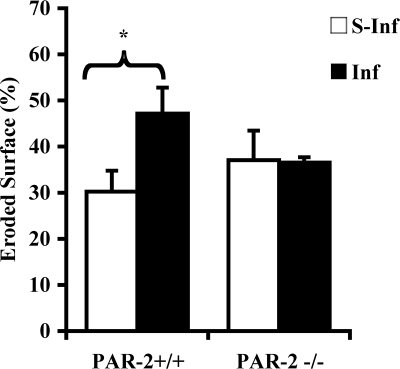
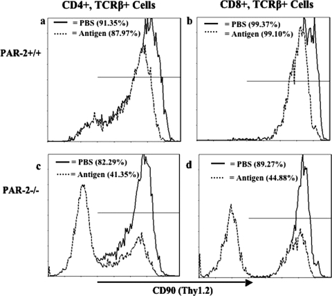
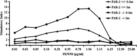
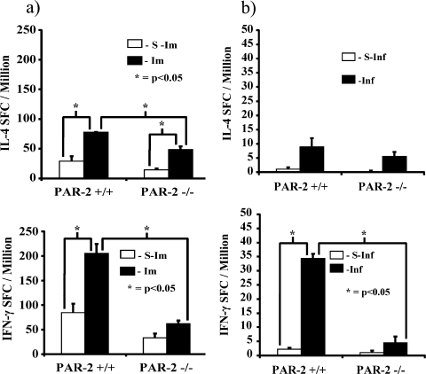
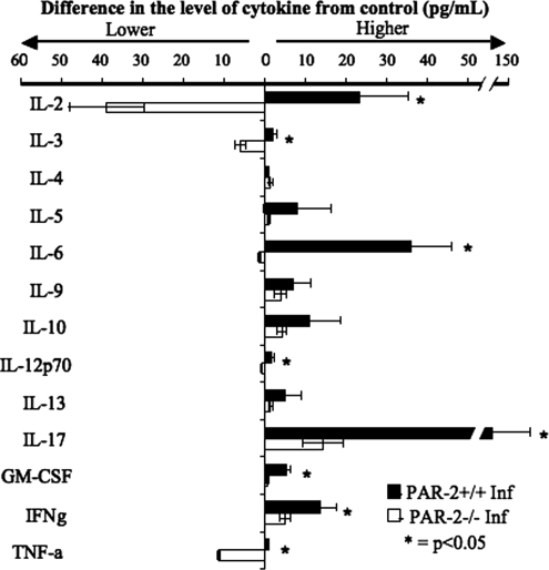
The tissue destruction seen in chronic periodontitis is commonly accepted to involve extensive upregulation of the host inflammatory response. Protease-activated receptor 2 (PAR-2)-null mice infected with Porphyromonas gingivalis did not display periodontal bone resorption in contrast to wild-type-infected and PAR-1-null-infected mice. Histological examination of tissues confirmed the lowered bone resorption in PAR-2-null mice and identified a substantial decrease in mast cells infiltrating the periodontal tissues of these mice. T cells from P. gingivalis-infected or immunized PAR-2-null mice proliferated less in response to antigen than those from wild-type animals. CD90 (Thy1.2) expression on CD4(+) and CD8(+) T-cell-receptor beta (TCRbeta) T cells was significantly (P < 0.001) decreased in antigen-immunized PAR-2-null mice compared to sham-immunized PAR-2-null mice; this was not observed in wild-type controls. T cells from infected or antigen-immunized PAR-2-null mice had a significantly different Th1/inflammatory cytokine profile from wild-type cells: in particular, gamma interferon, interleukins (interleukin-2, -3, and -17), granulocyte-macrophage colony-stimulating factor, and tumor necrosis factor alpha demonstrated lower expression than wild-type controls. The absence of PAR-2 therefore appears to substantially decrease T-cell activation and the Th1/inflammatory response. Regulation of such proinflammatory mechanisms in T cells and mast cells by PAR-2 suggests a pivotal role in the pathogenesis of the disease.


Figures

FIG. 1.
FIG. 1.
Regions used for histomorphometry in sections of mouse maxillae. (a) Section of a maxilla from a PAR-2+/+ mouse after challenge with P. gingivalis. The arrows indicate the fields in which the mast cells were counted. E, gingival epithelium; T, tooth; A, alveolar bone. (b) Section showing two mast cells (arrows); insert shows mast cells at higher magnification. (c) Section of a maxilla showing the field taken for measuring the eroded surface of the alveolar bone. The bold line indicates the region of interest, which is 450 μm in length. C, alveolar bone crest; T, tooth.
FIG. 2.
FIG. 2.
Quantification of periodontal bone loss, in PAR-1 (+/+ and −/−) and PAR-2 (+/+ and −/−) mice in experimental periodontitis. Half maxillae from PAR-2−/− (a) and PAR-2+/+ (b) mice orally infected with P. gingivalis W50 were stained with methylene blue. The area of exposed root surface (outlined by white lines in panel b) was measured and counted blind by one evaluator. The results for the comparison between infected and sham-infected PAR-1+/+ and PAR-1−/− (c) and PAR-2+/+ and PAR-2−/− (d) mice are shown as means ± SEM (n = 12; *, P < 0.05; **, P < 0.01).
FIG. 3.
FIG. 3.
Quantification of the number of mast cells in the periodontal tissue of mice orally challenged with P. gingivalis W50. The results are presented as the mean ± the SEM (n = 3). *, P < 0.05; **, P < 0.01 (for comparisons indicated by lines above the bars). S-Inf, sham-infected mice; Inf, infected mice.
FIG. 4.
FIG. 4.
Eroded bone surface measurements in sections of mouse maxillae (see Fig. 1c). The results are presented as means ± the SEM (n = 3; *, P < 0.05 for comparison between infected and sham-infected mice). S-Inf, sham-infected mice; Inf, infected mice.
FIG. 5.
FIG. 5.
Expression of CD90 on T cells of PAR-2+/+ and PAR-2−/− mice immunized with P. gingivalis FK-W50. Lymphocytes were isolated from inguinal and popliteal lymph nodes of mice 7 days after immunization. Expression of molecules on T-cell subsets was detected by FACS analysis with relevant antibodies. The data, expressed as cytometry histograms, are representative of three independent experiments. CD90 expression of T-cell (TCRβ+) subsets (CD4+ and CD8+) is represented by a solid line for PBS-immunized mice (control) and a dashed line for antigen (P. gingivalis FK-W50)-immunized mice for the PAR-2+/+ isolated lymphocytes (a and b) and for the PAR-2−/− isolated lymphocytes (c and d). A total of 10,000 events were measured.
FIG. 6.
FIG. 6.
T-cell proliferation assay. T cells isolated from the lymph nodes of mice 7 days after immunization were incubated with serially diluted P. gingivalis FK-W50 antigen in the presence of gamma-irradiated syngeneic antigen presenting cells in vitro. The T-cell proliferation was measured by determining the [3H]thymidine incorporation, with the results presented as the stimulatory index (SI), calculated by dividing the mean cpm obtained after antigen stimulation by the mean cpm detected in control, unstimulated wells. This graph is a representation of four independent experiments with similar results. S-Im, sham-immunized mice; Im, immunized mice.
FIG. 7.
FIG. 7.
Cytokine responses of P. gingivalis FK-W50-primed T cells from PAR-2+/+ and PAR-2−/− mice. T cells isolated from inguinal and popliteal lymph nodes of mice 7 days after immunization (a) and T cells isolated from submandibular lymph nodes (b) from the mouse periodontitis model were stimulated with P. gingivalis FK-W50 (1 μg/ml) in the presence of gamma-irradiated syngeneic antigen-presenting cells in vitro. After 2 days, the assay was stopped and developed. The data are expressed as SFC/million ± the SD minus the background and are the averages of triplicate assays. S-Im, sham-immunized mice; Im, immunized mice; S-Inf, sham-infected mice; Inf, infected mice.
FIG. 8.
FIG. 8.
Cytokine secretion responses of T cells from PAR-2+/+ and PAR-2−/− mice to stimulation with P. gingivalis FK-W50 antigen. T cells isolated from inguinal and popliteal lymph nodes of mice (PAR-2+/+ and PAR-2−/−) 7 days after immunization in the hind limb were stimulated with P. gingivalis FK-W50 in the presence of gamma-irradiated syngeneic antigen-presenting cells in vitro. Supernatants from the T-cell proliferation assays (Fig. 6) were collected, and supernatants corresponding to maximal T-cell proliferation in response to P. gingivalis FK-W50 antigen (0.78 μg/ml, Fig. 6) were analyzed for IL-2, IL-3, IL-4, IL-5, IL-6, IL-9, IL-10, IL-12p70, IL-13, IL-17, GM-CSF, IFN-γ, and TNF-α by using the Bio-Plex cytokine array system. The data are expressed in pg/ml minus the no-antigen control and are representative of three independent T-cell proliferation assays. Sample size, n = 3.

Similar articles

Cited by

References

    1. Abraham, L. A., C. Chinni, A. L. Jenkins, A. Lourbakos, N. Ally, R. N. Pike, and E. J. Mackie. 2000. Expression of protease-activated receptor-2 by osteoblasts. Bone 26:7-14. - PubMed
    1. Ahmed, Y. A., L. Tatarczuch, C. N. Pagel, H. M. Davies, M. Mirams, and E. J. Mackie. 2007. Physiological death of hypertrophic chondrocytes. Osteoarthritis Cartilage 15:575-586. - PubMed
    1. Alshurafa, H. N., G. R. Stenton, J. L. Wallace, M. D. Hollenberg, A. D. Befus, and H. Vliagoftis. 2004. A protease activated receptor-2 (PAR-2) activating peptide, tc-LIGRLO-NH2, induces protease release from mast cells: role in TNF degradation. BMC Pharmacol. 4:12-20. - PMC - PubMed
    1. Bacha, W., et al. 2000. Color atlas of veterinary histology, 2nd ed. Lippincott/The Williams & Wilkins Co., Baltimore, MD.
    1. Baggiolini, M., B. Dewald, and B. Moser. 1994. Interleukin-8 and related chemotactic cytokines—CXC and CC chemokines. Adv. Immunol. 55:97-179. - PubMed

Publication types

LinkOut - more resources