. 2010 Feb;30(3):694-710.
doi: 10.1128/MCB.00253-09. Epub 2009 Nov 23.

Cdk2 and Cdk4 regulate the centrosome cycle and are critical mediators of centrosome amplification in p53-null cells

Affiliations

Cdk2 and Cdk4 regulate the centrosome cycle and are critical mediators of centrosome amplification in p53-null cells

Arsene M Adon et al. Mol Cell Biol. 2010 Feb.

Erratum in

Abstract

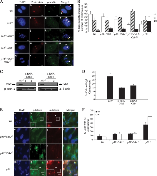
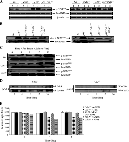
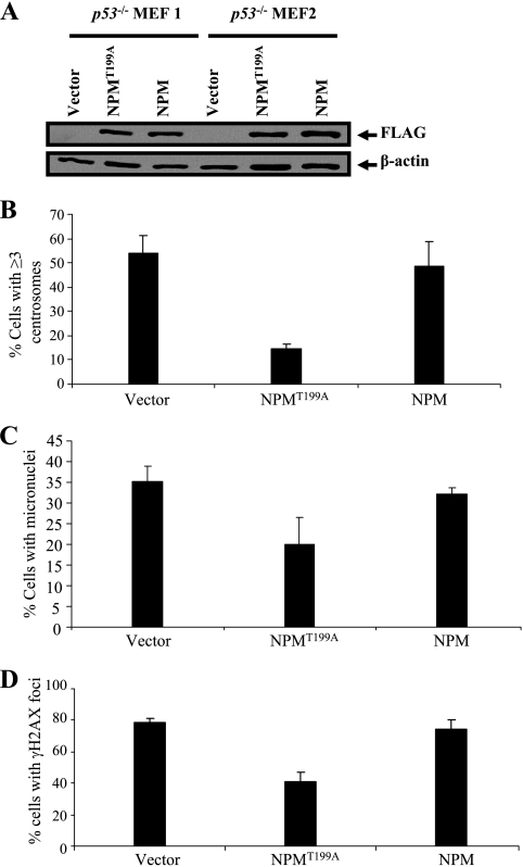
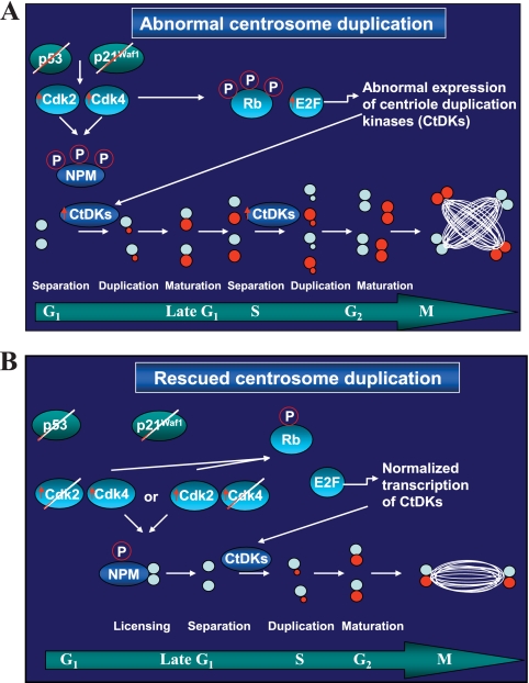
The two mitotic centrosomes direct spindle bipolarity to maintain euploidy. Centrosome amplification-the acquisition of > or =3 centrosomes-generates multipolar mitoses, aneuploidy, and chromosome instability to promote cancer biogenesis. While much evidence suggests that Cdk2 is the major conductor of the centrosome cycle and that it mediates centrosome amplification induced by various altered tumor suppressors, the role played by Cdk4 in a normal or deregulated centrosome cycle is unknown. Using a gene knockout approach, we report that Cdk2 and Cdk4 are critical to the centrosome cycle, since centrosome separation and duplication are premature in Cdk2(-)(/)(-) mouse embryonic fibroblasts (MEFs) and are compromised in Cdk4(-)(/)(-) MEFs. Additionally, ablation of Cdk4 or Cdk2 abrogates centrosome amplification and chromosome instability in p53-null MEFs. Absence of Cdk2 or Cdk4 prevents centrosome amplification by abrogating excessive centriole duplication. Furthermore, hyperactive Cdk2 and Cdk4 deregulate the licensing of the centrosome duplication cycle in p53-null cells by hyperphosphorylating nucleophosmin (NPM) at Thr199, as evidenced by observations that ablation of Cdk2, Cdk4, or both Cdk2 and Cdk4 abrogates that excessive phosphorylation. Since a mutant form of NPM lacking the G(1) Cdk phosphorylation site (NPM(T199A)) prevents centrosome amplification to the same extent as ablation of Cdk2 or Cdk4, we conclude that the Cdk2/Cdk4/NPM pathway is a major guardian of centrosome dysfunction and genomic integrity.


Figures

FIG. 1.
FIG. 1.
Genetic ablation of p53, Cdk2, or Cdk4 leads to absence of the respective protein expression. (A) PCR-based genotyping. Results of PCR analysis of genomic liver DNA from E.13.5 embryos generated by crossing p53+/ Cdk2+/ or p53+/ Cdk4+/ mice are shown. These gels included five double mutants (p53/ Cdk2/ [left panel] or p53/ Cdk4/ [right panel]), one Cdk2/ mutant, one Cdk4/ mutant, wild-type (Wt) embryos, and a control lacking DNA (H2O). M, molecular size marker; KO, knockout. (B) To confirm the genotyping data generated in panel A, Western blotting was performed using antibodies specific to p53, Cdk2, and Cdk4. β-Actin was used as a loading control (bottom). (C) Western blotting was conducted to determine the expression levels of Cdk4 in Cdk2/ MEFs and of Cdk2 in Cdk4/ MEFs. To ensure that equal amounts of proteins were loaded, β-actin was used to probe the same membrane.
FIG. 2.
FIG. 2.
Ablation of Cdk2 or Cdk4 does not significantly alter the cell cycle. (A and B) Cells of the indicated genotypes were arrested in G0 and subsequently stimulated by addition of serum. Cells were pulse-labeled with BrdU 30 min prior to harvest and were harvested at the indicated time points after serum stimulation. Cells were stained with anti-BrdU antibodies and the appropriate secondary antibodies and were visualized using confocal microscopy and a 63× objective. Nuclei were counterstained with DAPI. This experiment was repeated twice; results of a representative experiment are presented. Frequencies represent BrdU-positive cells in a population of at least 200 cells per group. Wt, wild type. (C) Whole-cell extracts were prepared from MEFs collected at the indicated time points following serum addition and were analyzed by Western blotting using antibodies against cyclin A, cyclin D1, cyclin E, and β-actin as a control. Cyc, cyclin. (D) Western blotting was performed with MEFs cultured in DMEM containing 0.2% FBS for 48 h. The numbers 1 and 2 above the lanes represent the loading of the protein lysates of two independent MEFs of the indicated genotypes. Western blots were probed with antibodies against p21Waf1, p27Kip1, p57Kip2, and p16INK4A. β-Actin served as a loading control. (E) Western blots of proteins extracted from controls (wild-type MEFs) or of wild-type MEFs transfected with siRNAs specific to p53 were probed with p53, p57, p16, and β-actin (control).
FIG. 3.
FIG. 3.
Ablation of Cdk2 and Cdk4 or siRNA-mediated silencing of Cdk2 and Cdk4 leads to distinct centrosome cycle defects. (A) Proliferating E13.5 mouse embryonic fibroblasts of the indicated genotypes were fixed, processed, and coimmunostained with anti-pericentrin, anti-γ-tubulin, and the appropriate secondary antibodies. Averages ± standard deviations of percentages of cells with one, two, and three centrosomes are shown. Exactly 8 wild-type (Wt), 3 Cdk2/, 4 Cdk4/, and 3 Cdk2/ Cdk4/ embryos were analyzed. The statistical significance of the averages (P ≤ 0.05) was established by an unequal-variance t test. t test values for the percentages of cells in each population containing one centrosome relative to that for the wild type were 0.159885 for Cdk2/ MEFs, 0.000518 for Cdk4/ MEFs, and 0.000544 for Cdk2/ Cdk4/ MEFs. The P values for the percentages of cells in each population containing two centrosomes relative to that of wild-type MEFs were 0.122182 for Cdk2/ MEFs, 0.000172 for Cdk4/ MEFs, and 0.000528 for Cdk2/ Cdk4/ MEFs. The P values for the percentages of cells with ≥3 centrosomes relative to that of wild-type MEFs were 0.091487 for Cdk2/ MEFs, 0.06122 for Cdk4/ MEFs, and 0.000808 for Cdk2/ Cdk4/ MEFs. (B) MEFs of the indicated genotypes were grown in duplicate to confluence, followed by serum starvation for 60 h. Quiescent cells were stimulated with serum, and every 4 h for a period of 16 h, the numbers of cells with one and two centrosomes were scored. This experiment was repeated twice; results of a representative experiment are presented. The BrdU data are the same as those presented in Fig. 2A and B; they are shown here for purposes of clarity. (C) Western blots of proteins extracted from nontransfected wild-type cells, or from cells transfected with siRNAs against Cdk2 or Cdk4, and probed with antibodies against Cdk2 or Cdk4. The same membrane was probed with β-actin as a control. (D) Wild-type MEFs that either were left untransfected or were transfected with siRNAs against Cdk2 or Cdk4 were immunostained with anti-γ-tubulin antibodies, and frequencies were established by counting cells with one and two centrosomes in a population of at least 200 cells per group. Three independent MEF groups were used. The statistical significance of the averages (P ≤ 0.05) was established by an unequal-variance t test. t test values for the percentage of cells in each population containing one centrosome relative to the percentage with two centrosomes were 0.215535 for wild-type MEFs, 0.003271 for MEFs transfected with a siRNA against Cdk2, and 0.008772 for those transfected with a siRNA against Cdk4. (E) Three independent Cdk4/ MEFs transfected with plasmids carrying either a control vector (pBABE-hygro) or pBABE-hygro-Cdk2 and plated after selection with hygromycin were immunostained using anti-γ-tubulin antibodies and the appropriate secondary antibodies. Frequencies were established by counting cells with one and two centrosomes in a population of at least 200 cells per group. t test values of the percentage of cells in each population containing one centrosome relative to the percentage with two centrosomes were 0.345271 for pBABE-hygro-transfected MEFs and 0.136406 for pBABE-hygro-Cdk2-transfected MEFs.
FIG. 4.
FIG. 4.
Ablation or siRNA-mediated silencing of Cdk2 and Cdk4 prevents centriole reduplication and centrosome amplification in p53/ MEFs. (A) MEFs of the indicated genotypes were coimmunostained with antibodies recognizing pericentrin (red) (b, f, j, n, and r) and γ-tubulin (green) (c, g, k, o, and s). Nuclei were stained with DAPI (blue) (a, e, I, m, and q). (d, h, l, p, and t) Overlay images of the pericentrin and γ-tubulin immunostaining. Wt, wild type. (B) Proliferating E13.5 mouse embryonic fibroblasts of the indicated genotypes were fixed, processed, and coimmunostained with anti-pericentrin, anti-γ-tubulin, and the appropriate secondary antibodies. The graph presents averages ± standard deviations of the percentages of cells with one, two, and three or more centrosomes. Exactly 8 wild-type, 8 p53/, 5 p53/ Cdk2/, 5 p53/ Cdk4/, and 4 p53/ Cdk2/ Cdk4/ embryos were analyzed. t test values for the percentage of cells in each population containing one centrosome (relative to that for the wild type) were 0.017174 for p53/ MEFs, 0.137854 for p53/ Cdk2/ MEFs, 0.358121 for p53/ Cdk4/ MEFs, and 3.95E-05 for p53/ Cdk2/ Cdk4/ MEFs. P values for the percentage of cells in each population containing two centrosomes relative to that for wild-type MEFs were 0.860687 for p53/ MEFs, 0.9713 for p53/ Cdk2/ MEFs, 0.024679 for p53/ Cdk4/ MEFs, and 2.69E-05 for p53/ Cdk2/ Cdk4/ MEFs. P values for the percentage of cells with ≥3 centrosomes relative to that for wild-type MEFs were 0.006967 for p53/ MEFs, 0.232114 for p53/ Cdk2/ MEFs, 0.706722 for p53/ Cdk4/ MEFs, and 0.051209 for p53/ Cdk2/ Cdk4/ MEFs. (C) Western blots of extracts from untransfected p53/ MEFs or p53/ MEFs transfected with Cdk2- or Cdk4-specific siRNAs were probed with the indicated primary antibodies. (D) Frequencies of centrosome amplification in control p53/ MEFs and in p53/ MEFs in which Cdk2 or Cdk4 was knocked down. Three independent MEFs were used. Centrosomes were detected as for panels A and B. The statistical significance of the averages (P ≤ 0.05) was established by an unequal-variance t test. P values for the percentage of cells with ≥3 centrosomes relative to that of control p53/ MEFs were 0.002445 for MEFs transfected with a Cdk2-specific SiRNA and 0.006696 for MEFs transfected with a Cdk4-specific siRNA. (E) Proliferating MEFs of the indicated genotypes (3 per group) were either left untreated (NT) or treated with 2 mM HU for 48 h. To determine the presence of centrioles, the cells were subjected to cold treatment and brief extraction prior to fixation. This treatment destabilizes microtubules nucleated at centrosomes; hence, centrioles can be microscopically visualized by immunostaining for α-tubulin (a major component of centrioles) at a high magnification. Cells were coimmunostained with anti-γ-tubulin polyclonal (green) (b, f, j, and n) and anti-α-tubulin monoclonal (red) (c, g, k, and o) antibodies and were counterstained with DAPI (blue) (a, e, i, and m). (d, h, l, and p) Overlaid images of γ-tubulin and α-tubulin immunostaining. (Insets) Magnified images of the areas indicated. (F) Frequencies of centriole reduplication were established by counting cells with ≥3 separated centrioles in a population of at least 200 cells per group. P values for HU-treated compared to NT cells were 0.791492 for wild-type MEFs, 1 for p53/ Cdk2/ MEFs, 0.507158 for p53/ Cdk4/ MEFs, and 0.012161 for p53/ MEFs.
FIG. 5.
FIG. 5.
Ablation of Cdk2 and Cdk4 inhibits chromosome instability in cells lacking p53. (A and B) The frequencies of γ-H2AX foci (arrows) in cells with the indicated genotypes were calculated. Bars, 10 μm. The graph (B) shows the average percentage of γ-H2AX foci in each population of at least 200 cells. Error bars, standard deviations. Each group included 4 different MEFs. P values (relative to the wild-type control) were 0.015218 for p53/ MEFs, 0.126173 for p53/ Cdk2/ MEFs, and 0.346771 for p53/ Cdk4/ MEFs. (C and D) Proliferating E13.5 mouse embryonic fibroblasts of the indicated genotypes were fixed, and nuclei were visualized with DAPI. Frequencies of micronucleus (insets and arrows) formation were calculated for at least 500 cells of each of the indicated genotypes. Each group included 4 different MEFs. P values (relative to the wild-type control) were 0.016122 for p53/ MEFs, 0.137054 for p53/ Cdk2/ MEFs, and 0.370282 for p53/ Cdk4/ MEFs.
FIG. 6.
FIG. 6.
Cdk2 and Cdk4 affect the centrosome cycle and centrosome amplification through NPM. (A) E13.5 MEFs of the indicated genotypes were serum starved for 60 h. Cells were preincubated with calyculin A, a serine/threonine phosphatase inhibitor. (Top) Western blot analysis of protein fractions of G0-arrested MEFs probed with antibodies against phospho-NPMT199 (p-NPMT199). (Center and bottom) Blots were probed with antibodies against total nucleophosmin and β-actin to show equal loading. (B) MEFs of the indicated genotypes were treated with 2 mM HU for 48 h and were then preincubated with calyculin A, a serine/threonine phosphatase inhibitor, before protein extraction. Western blots of the protein extracts were probed with antibodies against NPMT199 or against total NPM (control). The MEFs for the left panel are independent of those for the right. (C) Western blot analyses of MEFs of the indicated genotypes that were serum arrested and released into the cell cycle for various times. Western blots of the protein extracts were probed with antibodies against NPMT199 or against total NPM (control). (D) Western blots of Cdk4 immunoprecipitated (Ip-Cdk4) from extracts of wild-type, Cdk2/, or Cdk4/ MEFs were probed with antibodies against Cdk4 and cyclin D1. (E) Cdk4 kinase assays of protein lysates from wild-type, Cdk2/, and Cdk4/ MEFs were carried out at various time points following serum addition. Results for the 0-, 4-, and 8-h time points are shown. The results are from three independent MEFs. The experiment was repeated at least twice, and results of a representative experiment are presented. The reaction mixtures either contained NPM peptide (+ NPM) or contained no NPM peptide (no NPM). Luminescence was recorded by the SpectraMax Gemini XS luminometer using the SoftMax program. P values of kinase assays comparing NPM to no NPM at each indicated time point were 0.107203829 for lysates from wild-type MEFs at 0 h, 0.037437678 for those from Cdk2/ MEFs at 0 h, 0.000111335 for those from wild-type MEFs at 4 h, 0.002861355 for those from Cdk2/ MEFs at 4 h, 0.000761355 for those from wild-type MEFs at 8 h, and 0.000449084 for those from Cdk2/ MEFs at 8 h. The P values from the kinase assays performed with Cdk4/ MEFs were greater than 0.05 at any given time point.
FIG. 7.
FIG. 7.
NPMT199A suppresses centrosome amplification and chromosome instability. (A) Passage 2 p53/ MEFs were transiently transfected with plasmids encoding FLAG epitope-tagged NPM and the NPM/B23 mutant (NPMT99A). As a control, an empty vector was transfected. After neomycin selection, cell lysates were obtained and then probed with anti-FLAG antibodies. (B) The transfectants described in the legend to panel A were fixed, immunostained with anti-γ-tubulin polyclonal antibodies, and detected with Alexa Fluor 488-conjugated secondary antibodies. Cells were counterstained with DAPI. The number of cells with ≥3 centrosomes in a population of at least 200 cells was statistically analyzed by fluorescence microscopy. Each group included three transfected MEFs. P values (relative to the result for the transfected vector control) were 0.006963 for NPMT199A and 0.560677 for wild-type NPM. (C) Proliferating E13.5 mouse embryonic fibroblasts of the indicated genotypes were fixed, and nuclei were visualized with DAPI. The frequencies of micronucleus formation in a population of 500 cells were calculated for the indicated genotypes. P values (relative to the result for the transfected vector control) were 0.011338 for NPMT199A and 0.353737 for wild-type NPM. (D) Frequencies of γ-H2AX foci in cells with the indicated genotypes were calculated. P values (relative to the result for the transfected vector control) were 0.002591 for NPMT199A and 0.476327 for wild-type NPM.
FIG. 8.
FIG. 8.
Model explaining how the ablation of Cdks prevents centrosome amplification. (A) Ablation of p53 results in undetectable levels of p21Waf1, leading to hyperactive Cdk2 and Cdk4. Hyperactive Cdks cross talk to the centrosome in two modes. First, hyperactive Cdks hyperphosphorylate Rb in the nucleus, leading to uncontrolled E2F-dependent transcription of molecules that influence various steps in the centrosome duplication cycle: those involved in centriole splitting, as well as centriole duplication kinases (CtDKs). Second, hyperactive Cdks constitutively phosphorylate NPMT199, resulting in excessive licensing of centrosome duplication. Uncontrolled expression of CtDKs and the inability of NPM to suppress normal centrosome duplication lead to faster centrosome duplication cycles within a single cell cycle, resulting in the formation of multiple centrosomes. (B) When Cdk2 or Cdk4 is deleted from p53/ MEFs, Rb is underphosphorylated, and the E2F-dependent transcription of CtDKs is restored. In addition, underphosphorylated NPM restores normal centrosome licensing and prevents excessive centriole duplication. This restricts the centrosome duplication cycle to one per cell cycle, thus resulting in normal centrosome numbers.

References

    1. Albertson, D. G., C. Collins, F. McCormick, and J. W. Gray. 2003. Chromosome aberrations in solid tumors. Nat. Genet. 34:369-376. - PubMed
    1. Aleem, E., H. Kiyokawa, and P. Kaldis. 2005. Cdc2-cyclin E complexes regulate the G1/S phase transition. Nat. Cell Biol. 7:831-836. - PubMed
    1. Ball, K. L., S. Lain, R. Fahraeus, C. Smythe, and D. P. Lane. 1997. Cell-cycle arrest and inhibition of Cdk4 activity by small peptides based on the carboxy-terminal domain of p21WAF1. Curr. Biol. 7:71-80. - PubMed
    1. Bearss, D. J., R. J. Lee, D. A. Troyer, R. G. Pestell, and J. J. Windle. 2002. Differential effects of p21(WAF1/CIP1) deficiency on MMTV-ras and MMTV-myc mammary tumor properties. Cancer Res. 62:2077-2084. - PubMed
    1. Berthet, C., E. Aleem, V. Coppola, L. Tessarollo, and P. Kaldis. 2003. Cdk2 knockout mice are viable. Curr. Biol. 13:1775-1785. - PubMed

Publication types

MeSH terms