. 2009 Dec 15;183(12):8004-14.
doi: 10.4049/jimmunol.0901937.

In a murine tuberculosis model, the absence of homeostatic chemokines delays granuloma formation and protective immunity

Affiliations

In a murine tuberculosis model, the absence of homeostatic chemokines delays granuloma formation and protective immunity

Shabaana A Khader et al. J Immunol. .

Abstract

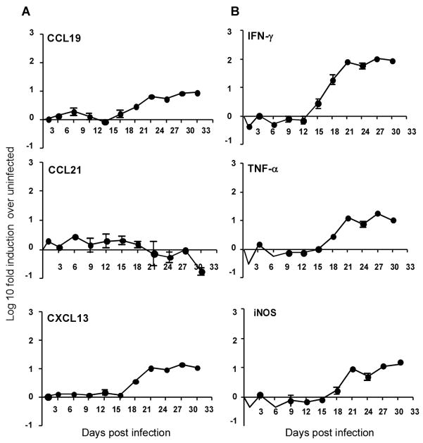
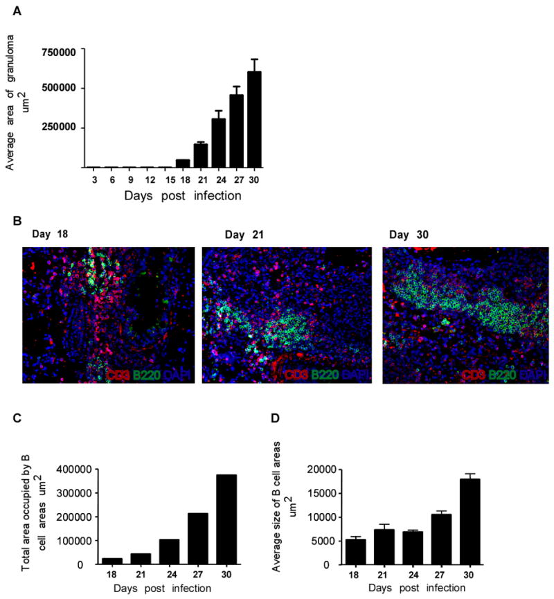
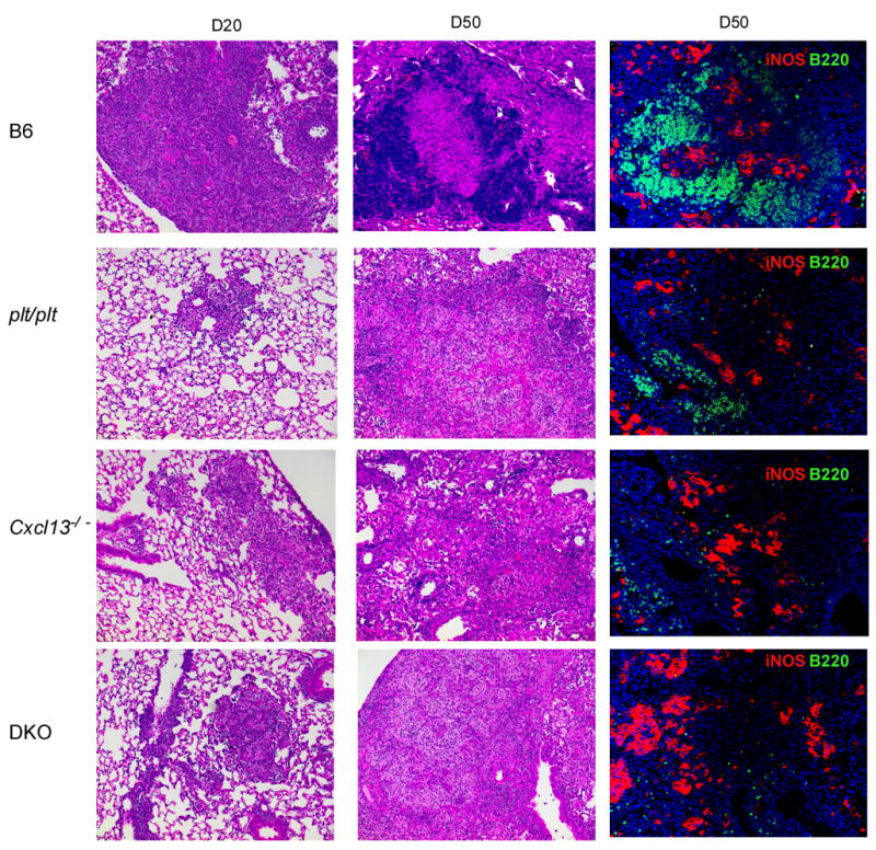
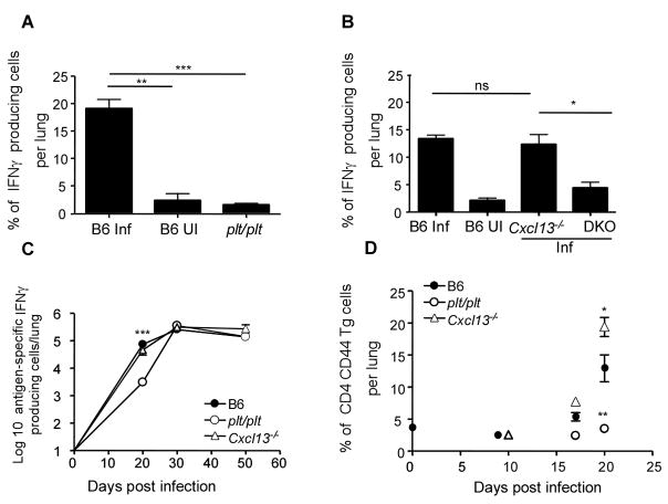
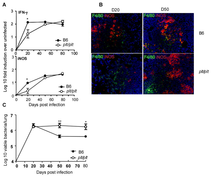
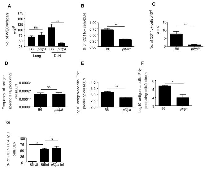
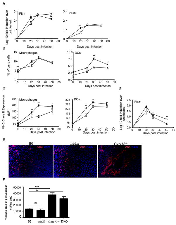
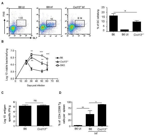
Mycobacterium tuberculosis infection (Mtb) results in the generation of protective cellular immunity and formation of granulomatous structures in the lung. CXCL13, CCL21, and CCL19 are constitutively expressed in the secondary lymphoid organs and play a dominant role in the homing of lymphocytes and dendritic cells. Although it is known that dendritic cell transport of Mtb from the lung to the draining lymph node is dependent on CCL19/CCL21, we show in this study that CCL19/CCL21 is also important for the accumulation of Ag-specific IFN-gamma-producing T cells in the lung, development of the granuloma, and control of mycobacteria. Importantly, we also show that CXCL13 is not required for generation of IFN-gamma responses, but is essential for the spatial arrangement of lymphocytes within granulomas, optimal activation of phagocytes, and subsequent control of mycobacterial growth. Furthermore, we show that these chemokines are also induced in the lung during the early immune responses following pulmonary Mtb infection. These results demonstrate that homeostatic chemokines perform distinct functions that cooperate to mediate effective expression of immunity against Mtb infection.


Figures

Figure 1
Figure 1. Homeostatic chemokines are induced in the lungs during the early immune response following Mtb infection
B6 mice were infected with ~75 CFU Mtb via the aerosol route and at specific times after infection, lung tissue was harvested and processed to extract RNA. The presence of mRNA for different homeostatic chemokines CCL19, CCL21 and CXCL13 (A) and protective molecules IFNγ, TNFα and iNOS (B) was determined by real-time PCR, and the log10 fold-increase in mRNA was determined for four infected mice vs four uninfected mice. The data points represent the mean and SD for four mice for each time point. One experiment representative of two shown.
Figure 2
Figure 2. Early induction of homeostatic chemokines correlate with the initiation of granuloma formation in the lung following Mtb infection
B6 mice were infected as described in Figure 1. Upon harvest, the caudal lobe of the lung from each of four mice per group was perfused with 10% formal saline, embedded in paraffin, sectioned, and stained using H&E. Morphometric analysis of area covered by the granuloma in H & E stained sections of Mtb-infected B6 lungs at different time points during the early immune response was determined(A). The data points represent the mean and SD for four mice for each time point. Lung sections from infected B6 mice at different time points post infection were stained for B220 (green) and CD3 (red) and shown at 40x magnification (B). Representative of four mice per group. Morphometric analysis of total area (C) and average area (D) covered by the B cell areas (B220+) in sections of infected lungs of B6 mice at different time points following infection. The data points represent the mean and SD for four mice for each time point. One experiment representative of two shown.
Figure 3
Figure 3. Expression of homeostatic chemokines is important for initiation and maintenance of granuloma formation following Mtb infection
B6 and chemokine deficient mice were infected as described for Figure 1 and lungs were fixed in 10% formalin, embedded in paraffin and stained using H &E as described in Figure 2. The panels in the left column show infected lungs (x10) at day 20 post infection. The panels in the middle column show the cellular components of the granulomata at day 50 post infection (x10). Areas of lymphocyte accumulation appear as darker areas, while areas of macrophage accumulation appear lighter. The panels in the right column show immunofluorescence for B cells (B220 in green) and iNOS (red) on day 50 post infection (x20). One experiment representative of two is shown, n = 4 mice per group.
Figure 4
Figure 4. Delayed accumulation of IFNγ producing T cells in the absence of CCL19/CCL21 but not CXCL13
B6 (B6 Inf) and plt/plt mice (A) or B6 (B6 Inf), Cxcl13−/− and DKOs (B) were infected as in Figure 1. B6 uninfected controls are also shown (B6 UI)(A,B). Lymphocytes were isolated from the lung at day 20 post infection and were cultured with PMA/Ionomycin in the presence of golgi stop for 5 hours. The presence of intracellular IFNγ in CD4 T lymphocytes was determined by flow cytometry (A,B). Cells from infected B6 (closed circles), plt/plt (open circles), Cxcl13−/− (open triangles) were cultured with irradiated feeder cells and ESAT 6 peptide for 24 h and the number of IFN-γ producing antigen-specific cells determined by ELISpot(C). 1×10⁶ ESAT-6 TCR Tg CSFE labeled naïve T cells were transferred into infected B6 (closed circles), plt/plt (open circles) or Cxcl13−/−(open triangles) mice at different time points post infection (D). The expression of CD44 on transferred Tg T cells was determined 12 hours later. The data points represent the mean (±SD) of values from four-five mice (A, B, C, and D). ﹡, p ≤0.05. ﹡﹡, p ≤0.005. ﹡﹡﹡, p ≤0.0005. ns-not significant.
Figure 5
Figure 5. Delay in the generation of protective acquired responses in plt/plt mice results in establishment of higher bacterial numbers
B6 (closed circles) and plt/plt mice (open circles) were infected as described for Figure 1 and at various times post infection lung tissue was harvested and processed to extract RNA (A). The presence of specific mRNA was determined by real-time PCR and the log10-fold-increase determined for three to four infected mice over four uninfected mice. The data points represent the mean and SD values for four mice for each time point. The expression of iNOS (red) by macrophages expressing F4/80 (green) in lungs from Mtb infected B6 and plt/plt mice was determined at day 20 and day 50 post infection (B). B6 (closed circles) and plt/plt (open circles) mice were infected via the aerosol route with ~ 75 M. tuberculosis H37Rv bacteria. The bacterial burden was determined in the lungs of B6 and plt/plt infected mice over time(C). The graph shows one experiment representative of two. The data points represent the mean (±SD) of values from four-five mice (A, C). ﹡, p ≤0.05. ﹡﹡, p ≤0.005.
Figure 6
Figure 6. Compromised generation of IFNγ producing T cells in the absence of CCL19/CCL21
B6 and plt/plt mice were infected as described for Figure 1. Cells from wild type and plt/plt infected lungs and draining lymph nodes were isolated at day 20 and total numbers of WBCs were determined per organ (A). The frequency(B) of CD11c+ cells in the lymph nodes of B6 and plt/plt mice were determined by flow cytometry and total numbers of CD11c+ cells was determined(C). The frequency (D) and total numbers (E,F) of IFNγ producing antigen specific cells were determined by ESAT-6 driven ELISpot assay in draining lymph nodes (E) and spleen (F). 1×10⁶ ESAT-6 TCR Tg CSFE labeled naïve T cells were transferred into infected B6 and plt/plt mice at day 20 post infection and the lymph nodes were harvested and processed to generate single cell suspensions 12 hours later(F). The expression of CD69 was determined on transferred Tg CD4 T cells using flow cytometry. The data points represent the mean (±SD) of values from four-five mice (A–F). ﹡, p ≤0.05. ﹡﹡, p ≤0.005, ns-not significant.
Figure 7
Figure 7. CXCL13 is required for spatial organization of lymphocytes within the granulomas
B6 (closed circles) and Cxcl13−/− mice (open triangles) were infected as described for Figure 1 and at specific times post infection lung tissue was harvested and processed to extract RNA (A). The presence of specific mRNA was determined by real-time PCR and the log10-fold-increase in mRNA determined for three to four infected mice vs four uninfected mice. The data points represent the mean and SD values for four mice for each time point. Expression of MHC Class II was determined on CD11b+ and CD11c+ cells isolated from lungs of infected B6 (filled circles) or Cxcl13−/− mice (open traingles) on day 20 post infection(B). The presence of Fizz 1 mRNA was determined by real-time PCR and the log10-fold-increase in mRNA determined for three to four infected mice vs four uninfected mice (C). The distribution of CD3 lymphocytes in lungs of infected B6, plt/plt and Cxcl13−/− mice was determined by immunofluorescence on day20 (D) and the area occupied by CD3 T lymphocytes in the perivascular cuffs was determined morphometrically (E).
Figure 8
Figure 8. CXCL13 is required for recruitment of GC B cells and control of mycobacteria
B6 and Cxcl13−/− mice were infected as described for Figure 1 and the frequency of CD19/B220 positive B cells expressing GL7 and CD95/FAS (GC B cells) from lungs of infected B6 and Cxcl13−/− mice was determined by flow cytometry at day 20 post infection (A). B6 uninfected controls are also shown (B6 UI). B6 (closed circles), Cxcl13−/− (open triangles) and DKO (open squares) were infected via the aerosol route with 75 M. tuberculosis H37Rv bacteria and the bacterial burden determined in the lungs over time(B). B6 and Cxcl13−/− mice were infected as described for Figure 1. Cells from wild type and Cxcl13−/− infected spleens were isolated at day 20 and the number of antigen-specific IFNγ-producing cells determined by ELISpot (C). Uninfected (B6 UI) and day 20-infected B6 and Cxcl13−/− mice received CFSE labeled ESAT-6 TCR Tg T cells and the frequency of Tg cells expressing CD69 12 hours after transfer was determined in the spleens by flow cytometry (D). The data points represent the mean (±SD) of values from four-five mice (A–F). ﹡, p ≤0.05. ﹡﹡, p ≤0.005. ﹡﹡﹡, p ≤0.0005, ns-not significant.

References

    1. Flynn J, Chan J. Immunology of tuberculosis. Annual Review of Immunology. 2001;19:93–129. - PubMed
    1. Bean AG, Roach DR, Briscoe H, France MP, Korner H, Sedgwick JD, Britton WJ. Structural deficiencies in granuloma formation in TNF gene-targeted mice underlie the heightened susceptibility to aerosol Mycobacterium tuberculosis infection, which is not compensated for by lymphotoxin. J Immunol. 1999;162:3504–3511. - PubMed
    1. Cyster JG. Chemokines and cell migration in secondary lymphoid organs. Science. 1999;286:2098–2102. - PubMed
    1. Cyster JG. Chemokines and the homing of dendritic cells to the T cell areas of lymphoid organs. J Exp Med. 1999;189:447–450. - PMC - PubMed
    1. Gunn MD, Tangemann K, Tam C, Cyster JG, Rosen SD, Williams LT. A chemokine expressed in lymphoid high endothelial venules promotes the adhesion and chemotaxis of naive T lymphocytes. Proc Natl Acad Sci U S A. 1998;95:258–263. - PMC - PubMed

Publication types

MeSH terms