Comparative Study
. 2009 Dec 15;183(12):7984-93.
doi: 10.4049/jimmunol.0901336.

Tumor progression locus 2 (Map3k8) is critical for host defense against Listeria monocytogenes and IL-1 beta production

Affiliations
Comparative Study

Tumor progression locus 2 (Map3k8) is critical for host defense against Listeria monocytogenes and IL-1 beta production

Lisa A Mielke et al. J Immunol. .

Abstract

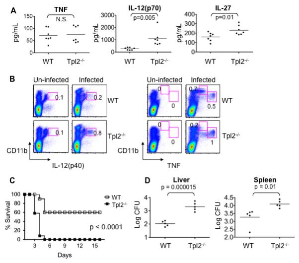
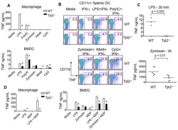
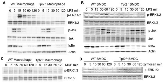
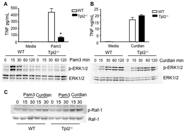
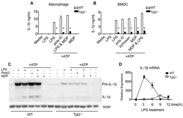
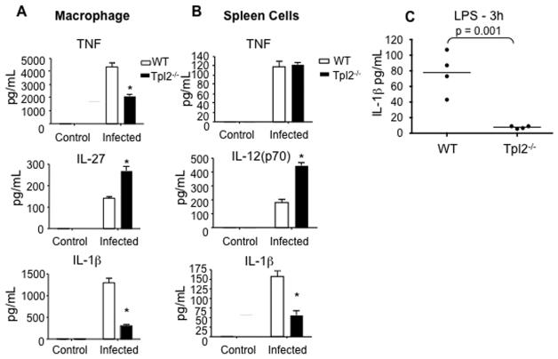
Tumor progression locus 2 (Tpl2, also known as Map3k8 and Cot) is a serine-threonine kinase critical in innate immunity, linking toll-like receptors (TLRs) to TNF production through its activation of ERK. Tpl2(-/-) macrophages have abrogated TNF production but overproduce IL-12 in response to TLR ligands. Despite enhanced IL-12 production, Tpl2(-/-) T cells have impaired IFN-gamma production. Therefore, the role of Tpl2 in a bona fide bacterial infection where all of these cytokines are important in host defense is unclear. To address this issue, we infected Tpl2(-/-) mice with the model pathogen Listeria monocytogenes. We found that Tpl2(-/-) mice infected i.v. with L. monocytogenes had increased pathogen burdens compared with wild-type mice and rapidly succumbed to infection. Enhanced susceptibility correlated with impaired signaling through TLR2 and nucleotide-binding oligomerization domain 2, two receptors previously shown to mediate Listeria recognition. Surprisingly, TNF production in response to infection was not significantly impaired, even though Tpl2 has been implicated in the regulation of TNF. We found that the role of Tpl2 has cell-type specific effects in regulating TNF and transduces signals from some, but not all, pattern recognition receptors (PRR). In contrast to the cell-type- and receptor-specific regulation of TNF, we found that Tpl2 is essential for IL-1beta production from both macrophages and dendritic cells. These studies implicate Tpl2 as an important mediator for collaboration of pattern recognition receptors with danger-associated molecular patterns to induce TNF and IL-1beta production and optimal host defense.


Figures

Figure 1
Figure 1. Tpl2 is important for host defense against Listeria monocytogenes
(A) Normal TNF production in serum of Tpl2-deficient mice infected with Listeria monocytogenes. Wild-type (WT) and Tpl2−/− mice were infected i.v. with 2×10³ CFU L. monocytogenes for 48 h. Sera were collected and analyzed for TNF, IL-12(p70) and IL-27. (B) L. monocytogenes-induced TNF production is dependent on Tpl2 in some, but not all innate immune cells. Spleens were removed from infected mice and splenocytes were fixed and permeabilized. Intracellular cytokine levels were assessed by flow cytometry. (C) Tpl2-deficient mice have enhanced lethality in response to L. monocytogenes. WT and Tpl2−/− mice were infected i.v. with 2×10⁴ CFU L. monocytogenes, and survival was monitored. (D) Lethality in Tpl2−/− mice results from impaired host defense. WT and Tpl2−/− mice were infected i.v. with 2×10³ CFU L. monocytogenes for 48 h. Spleens and livers were homogenized and bacterial burden was measured by plating serial dilutions of homogenates on LB agar plates. Results are representative of 3 – 4 independent experiments. Error bars correspond to standard deviations and * denotes p<0.05 as determined by Student's t-test. Abbreviations: not significant (N.S.).
Figure 2
Figure 2. Tpl2 regulates TNF production in a cell type- and PRR-specific manner
(A) Tpl2 is required for TLR-induced TNF production in macrophages. Bone marrow derived macrophages or BMDCs from WT or Tpl2−/− mice were stimulated with LPS, poly IC, zymosan, R848 or CpG for 18 h, and TNF was measured in cell supernatants. (B) Tpl2 is not required for TNF production by splenic DCs. Spleen cells from WT or Tpl2−/− animals were cultured with IFN-γ or IFN-γ plus LPS, poly IC, zymosan, R848 or CpG for 4 h. Cells were stained with anti- CD11c and CD11b antibodies, fixed, permeabilized and stained with anti-TNF antibody. Dot plots are gated on CD11c+ cells. (C) Tpl2 controls LPS and zymosan-induced TNF production in vivo. WT or Tpl2−/− mice were injected intraperitoneally with LPS (30 min) or zymosan (3h), sera were collected to measure TNF concentrations. (D) Tpl2 is important for Nod2 signaling in both macrophages and BMDCs. Bone marrow derived macrophages were stimulated with LPS, MDP or combinations of LPS and MDP for 18 h. BMDCs were stimulated with LPS, zymosan, MDP or combinations of these ligands for 18 h, and TNF was measured in cell supernatants. Results are representative of 3 independent experiments. Error bars correspond to standard deviations and * denotes p<0.05 as determined by Student's t-test.
Figure 3
Figure 3. Tpl2 controls the MAP kinase and NF-κB pathways in a cell type-specific and stimulus-specific manner
(A-B) Enhanced activation of IκBα in Tpl2-deficient DCs. WT or Tpl2−/− (A) macrophages or (B) BMDCs were stimulated with LPS for 5, 15, 30, 60, or 120 min. Cell lysates were immunoblotted with antibodies against phospho-ERK1/2, phospho-Jnk or IκBα. Membranes were re-probed with antibodies against ERK1/2, Jnk or actin as loading controls. (C) Tpl2 links the Nod2 receptor to the ERK pathway. WT or Tpl2−/− macrophages were stimulated with MDP for 5, 15, 30, 60, or 120 min. The cell lysates were immunoblotted with anti-phospho-ERK1/2 or anti-ERK1/2. (D) Tpl2 is not required for zymosan-induced ERK activation. WT or Tpl2−/− BMDCs were stimulated with zymosan for the indicated times and cell lysates were probed with antibodies for phospho-ERK1/2. The membrane was stripped and re-probed for total ERK1/2 as a loading control. Results are representative of at least 2 independent experiments.
Figure 4
Figure 4. ITAM-containing PRRs can utilize a distinct MAP3K8
(A) Tpl2 is required for normal TNF production and ERK activation downstream of TLR2. WT or Tpl2−/− BMDCs were stimulated with Pam3 for 18 h to measure supernatant TNF levels. BMDCs were also stimulated with Pam3 for 15, 30, 60, or 120 min, cell lysates were probed for phospho-ERK1/2 and total ERK1/2. (B) Tpl2 is not required for dectin-1-induced TNF production or signal transduction. WT or Tpl2−/− BMDCs were stimulated with curdlan for 18h to measure supernatant TNF levels. Cells were also stimulated with curdlan for 15, 30, 60, or 120 min, western blot was performed and cell lysates were probed for phospho-ERK1/2 and total ERK1/2. (C) Raf-1 is activated in response to stimulation of dectin-1. Western blot was also performed on WT BMDCs stimulated with Pam3 or Curdlan for 15 or 30 min, cell lysates were probed with anti-phospho-Raf-1, the membrane was stripped and re-probed for total Raf-1 protein as a loading control. Results are representative of 2-3 independent experiments. Error bars are equal to standard deviation and * p<0.05 as determined by Student's t-test.
Figure 5
Figure 5. Tpl2 regulates IL-1β production in response to TLR stimulation
(A-B) Tpl2 is essential for IL-1β production by macrophages and BMDCs. (A) Macrophages from WT or Tpl2−/− mice were cultured and stimulated with LPS, poly IC or MDP and the combination of LPS and MDP for 4 h. The cells were stimulated with ATP for 20 min, after which IL-1β was measured in cell supernatants. (B) WT or Tpl2−/− BMDCs were stimulated with LPS, poly IC, zymosan, MDP separately and LPS in combination with MDP for 4 h, followed by a second stimulation with ATP for 20 min. Cell supernatants were collected, and IL-1β was measured. (C) Tpl2 is critical for production of pro-IL-1β protein. Macrophages were stimulated as in (A) cells were lysed and western blotting was performed to measure IL-1β protein. (D) Tpl2 is essential for induction of IL-1β mRNA. WT or Tpl2−/− bone marrow macrophages were stimulated with LPS for 3, 6, 9 and 12 h. The cells were lysed and IL-1β mRNA expression was analyzed by real-time PCR relative to a GADPH control. Error bars indicate standard deviations and * denotes p<0.05 as determined by Student's t-test.
Figure 6
Figure 6. Tpl2 controls IL-1β production in response to L. monocytogenes
(A-B) Tpl2 is important for IL-1β production by macrophages and spleen cells. Bone marrow derived macrophages (A) or total spleen cells (B) from WT or Tpl2−/− mice were infected with L. moncytogenes at a ratio of 1 bacteria to 40 cells for 2 h. Cells were treated with gentamicin, washed with fresh media, cultured for an additional 18 h and cytokines were measured in cell supernatants. (C) Tpl2 is important in vivo for the production of IL-1β. WT and Tpl2−/− mice were injected i.p with LPS. Serum was collected 3 h later, and IL-1β was measured.

References

    1. O'Neill LA. When signaling pathways collide: positive and negative regulation of toll-like receptor signal transduction. Immunity. 2008;29:12–20. - PubMed
    1. Akira S, Uematsu S, Takeuchi O. Pathogen recognition and innate immunity. Cell. 2006;124:783–801. - PubMed
    1. Ogura Y, Inohara N, Benito A, Chen FF, Yamaoka S, Nunez G. Nod2, a Nod1/Apaf-1 family member that is restricted to monocytes and activates NF-kappaB. J Biol Chem. 2001;276:4812–4818. - PubMed
    1. Girardin SE, Boneca IG, Viala J, Chamaillard M, Labigne A, Thomas G, Philpott DJ, Sansonetti PJ. Nod2 is a general sensor of peptidoglycan through muramyl dipeptide (MDP) detection. J Biol Chem. 2003;278:8869–8872. - PubMed
    1. Inohara N, Ogura Y, Fontalba A, Gutierrez O, Pons F, Crespo J, Fukase K, Inamura S, Kusumoto S, Hashimoto M, Foster SJ, Moran AP, Fernandez-Luna JL, Nunez G. Host recognition of bacterial muramyl dipeptide mediated through NOD2. Implications for Crohn's disease. J Biol Chem. 2003;278:5509–5512. - PubMed

Publication types

MeSH terms