. 2009 Dec 15;183(12):7919-30.
doi: 10.4049/jimmunol.0803903.

The yellow fever virus vaccine induces a broad and polyfunctional human memory CD8+ T cell response

Affiliations

The yellow fever virus vaccine induces a broad and polyfunctional human memory CD8+ T cell response

Rama S Akondy et al. J Immunol. .

Abstract

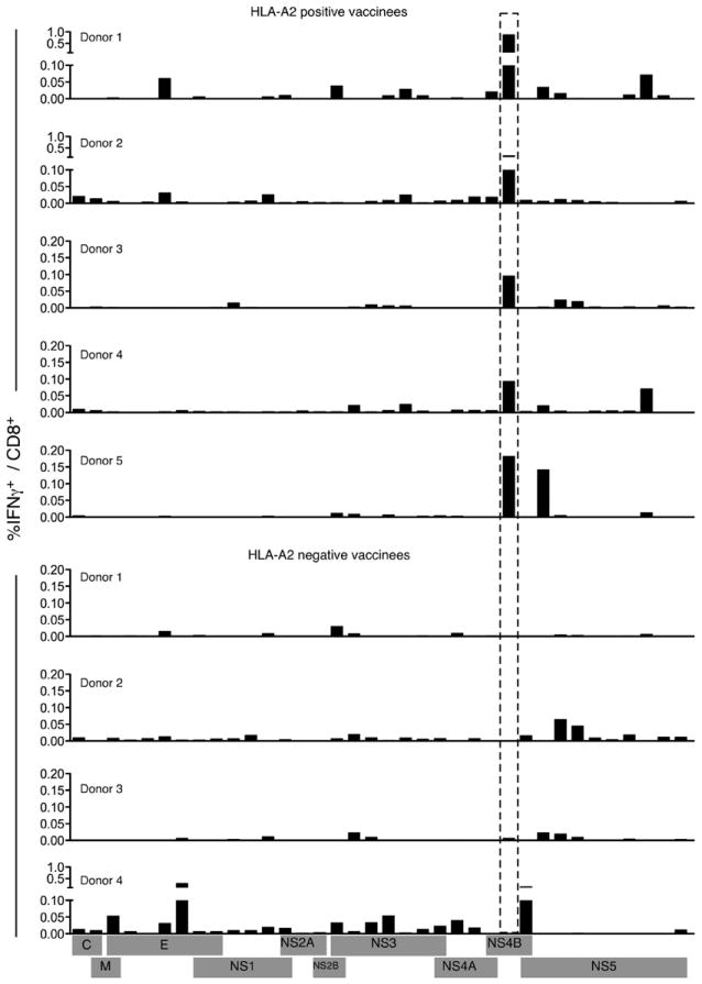
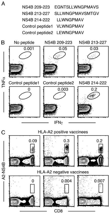
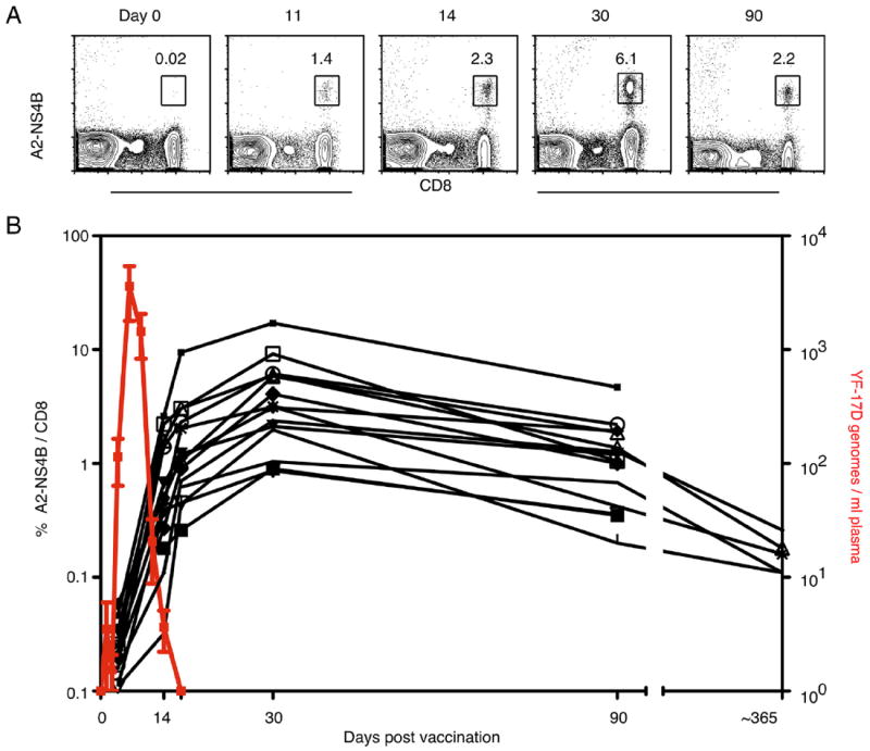
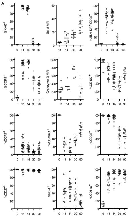
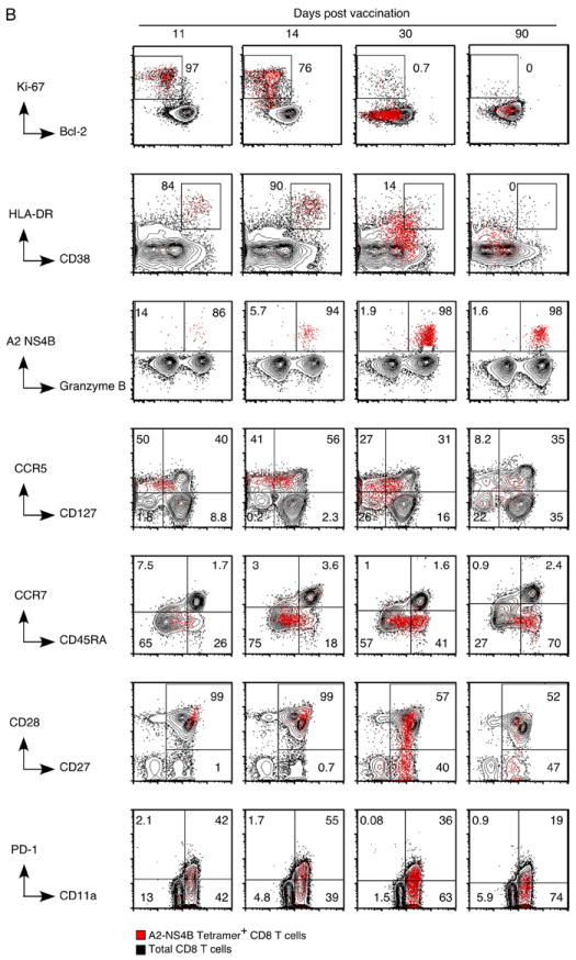
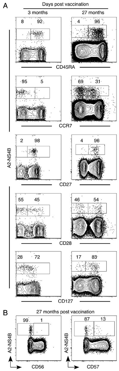
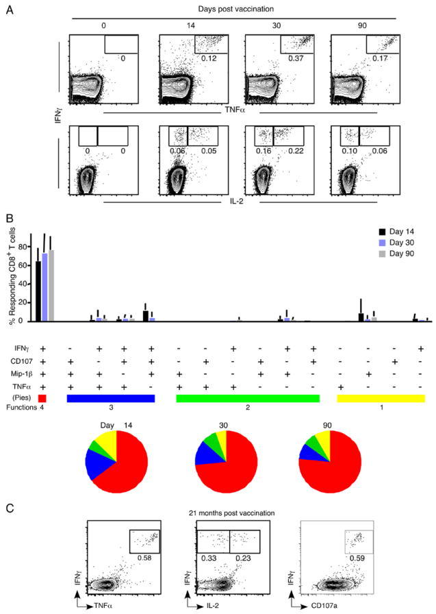
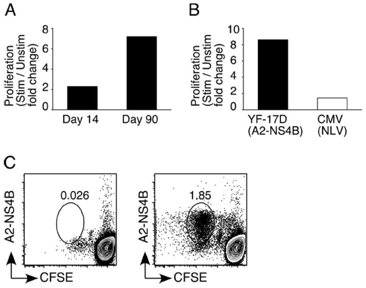
The live yellow fever vaccine (YF-17D) offers a unique opportunity to study memory CD8(+) T cell differentiation in humans following an acute viral infection. We have performed a comprehensive analysis of the virus-specific CD8(+) T cell response using overlapping peptides spanning the entire viral genome. Our results showed that the YF-17D vaccine induces a broad CD8(+) T cell response targeting several epitopes within each viral protein. We identified a dominant HLA-A2-restricted epitope in the NS4B protein and used tetramers specific for this epitope to track the CD8(+) T cell response over a 2 year period. This longitudinal analysis showed the following. 1) Memory CD8(+) T cells appear to pass through an effector phase and then gradually down-regulate expression of activation markers and effector molecules. 2) This effector phase was characterized by down-regulation of CD127, Bcl-2, CCR7, and CD45RA and was followed by a substantial contraction resulting in a pool of memory T cells that re-expressed CD127, Bcl-2, and CD45RA. 3) These memory cells were polyfunctional in terms of degranulation and production of the cytokines IFN-gamma, TNF-alpha, IL-2, and MIP-1beta. 4) The YF-17D-specific memory CD8(+) T cells had a phenotype (CCR7(-)CD45RA(+)) that is typically associated with terminally differentiated cells with limited proliferative capacity (T(EMRA)). However, these cells exhibited robust proliferative potential showing that expression of CD45RA may not always associate with terminal differentiation and, in fact, may be an indicator of highly functional memory CD8(+) T cells generated after acute viral infections.


Conflict of interest statement

Disclosures

The authors have no financial conflict of interest.

Figures

FIGURE 1
FIGURE 1
YF-17D elicits a broad diversity of memory CD8+ T cells. PBMC from YF-17D-vaccinated individuals were stimulated with peptide pools derived from YF-17D proteins and IFN-γ production was assayed by ICC. The percentage of IFN-γ+ CD8+ T cells for individual vaccinees is shown.
FIGURE 2
FIGURE 2
YF-17D harbors a dominant HLA-A2 restricted epitope in the NS4B protein. A, Sequences of the peptides used to confirm and identify the immunogenic nonamer epitope in NS4B. B, PBMC from HLA-A2+ vaccinees were cultured in the presence or absence of 15-mer peptides (NS4B 209–223 or NS4B213–227) and ICC staining was performed (upper panels). To confirm the nonamer epitope in these peptides, PBMC were stimulated with either the NS4B 214–222 peptide or with control peptides that differed by one amino acid (lower panels). Plots gated on CD8+ T cells for one representative donor are shown. C, Identification of YF-17D-specific CD8+ T cells using MHC class I tetramers. Peripheral blood from HLA-A2+ or HLA-A2 individuals vaccinated with YF-17D 2 wk previously was stained with the A2-NS4B tetramer. Plots are gated on all lymphocytes and numbers indicate the percentage of tetramer+ cells from total lymphocytes.
FIGURE 3
FIGURE 3
Kinetics of the YF-17D-specific primary CD8+ T cell response tracked by MHC class I tetramers. A, Tetramer kinetics in one representative vaccinee is shown. Plots show all lymphocytes but the numbers indicate the percentage of A2-NS4B+ cells from total CD8+ T cells. B, YF-17D genomes in the plasma (red line; mean ± SD) and the percentage of A2-NS4B+ (black lines; each for one vaccinee) in YF-17D vaccinees over 1 year is shown.
FIGURE 4
FIGURE 4
Effector and memory differentiation of YF-17D-specific CD8+ T cells. A, Results from the longitudinal phenotypic analysis of A2-NS4B+ cells present in blood of 10 to 15 vaccinees are summarized. The day 11, 14, 30, and 90 data are from the same group of vaccinees. The day 0 data are from a separate group and represent expression of the relevant marker on naive (CD45RA+CCR7+) CD8+ T cells. The percentage of A2-NS4B+ CD8+ T cells expressing the relevant marker for each individual (open circles) and the group mean (horizontal line) are shown. MFI, Mean fluorescence intensity. B, Flow plots representing the phenotype of YF-17D-specific CD8+ T cells. Plots are gated on total CD8+ T cells (black background) or YF-17D-specific (A2-NS4B+) CD8+ T cells (red dots). Numbers show the percentage of A2-NS4B+ CD8+ T cells in the gate.
FIGURE 4
FIGURE 4
Effector and memory differentiation of YF-17D-specific CD8+ T cells. A, Results from the longitudinal phenotypic analysis of A2-NS4B+ cells present in blood of 10 to 15 vaccinees are summarized. The day 11, 14, 30, and 90 data are from the same group of vaccinees. The day 0 data are from a separate group and represent expression of the relevant marker on naive (CD45RA+CCR7+) CD8+ T cells. The percentage of A2-NS4B+ CD8+ T cells expressing the relevant marker for each individual (open circles) and the group mean (horizontal line) are shown. MFI, Mean fluorescence intensity. B, Flow plots representing the phenotype of YF-17D-specific CD8+ T cells. Plots are gated on total CD8+ T cells (black background) or YF-17D-specific (A2-NS4B+) CD8+ T cells (red dots). Numbers show the percentage of A2-NS4B+ CD8+ T cells in the gate.
FIGURE 5
FIGURE 5
The YF-17D-specific CD8+ T cell memory is fine-tuned over 2 years. A, Comparison of the phenotype YF-17D-specific memory CD8+ T cells 3 mo and 2 years after vaccination. Representative flow plots (gated on total CD8+ T cells) from one of three vaccinees are shown. B, Representative flow plots (gated on total CD8+ T cells) show expression of the late differentiation markers CD56 and CD57 on YF-17D-specific memory CD8+ T cells in an individual vaccinated 2 years earlier.
FIGURE 6
FIGURE 6
YF-17D elicits polyfunctional, long-lived memory CD8+ T cells. A, Kinetics of cytokine-producing CD8+ T cells in individuals immunized with YF-17D. PBMC were isolated from vaccinees and responses to the NS4B 214–222 peptide were measured by ICC staining. Representative flow plots (gated on total CD8+ T cells) from one vaccinee are shown. B, Functional profile of the YF-17D-specific CD8+ T cell response. All possible combinations of four functions (degranulation by CD107a staining and secretion of IFN-γ, TNF-α, and Mip-1β) are shown on the x-axis. Bars indicate the percentage of the total response contributed by CD8+ T cells with the combination of responses on the x-axis. Responses are grouped according to the number of functions and the data summarized by the pie charts. Each slice of the pie represents the fraction of the total response that consists of CD8+ T cells positive for a given number of functions. Mean data from four YF-17D-immunized individuals are shown. C, Functional profile of the YF-17D-specific memory CD8+ T cells 2 years after vaccination. Plots gated on CD8+ T cells show the cytokines produced in response to the stimulation of PBMC with the NS4B 214–222 peptide. Data from one representative vaccinee are shown.
FIGURE 7
FIGURE 7
YF-17D elicits memory CD8+ T cells with a robust proliferative potential. A, Proliferative potential of YF-17D-specific effector (day 14) and memory (day 90) CD8+ T cells. PBMC from vaccinated individuals were labeled with CFSE and stimulated (Stim) or not (Unstim) with the A2-NS4B peptide for 6 days. CFSE dilution and staining with the A2-NS4B tetramer was used to identify divided cells. The percentage of CD8+ T cells that are A2-NS4B+ cells is shown as the fold increase above the percentage in blood. B, Expansion of YF-17D-specific memory CD8+ T cells compared with the expansion of CMV-specific CD8+ T cells from the same individual. CFSE-labeled PBMC were stimulated with either A2-NS4B or the CMV-NLV peptide for 6 days. The percentage of CD8+ T cells that are tetramer+ is shown as the fold increase above the percentage in blood. C, Retention of proliferation potential by YF-17D-specific memory CD8+ T cells. PBMC from an individual vaccinated with YF-17D 10 years previously were labeled and stimulated as described above and A2-NS4B specific responses were measured. Plots are gated on CD8+ T cells.

References

    1. Ahmed R, Gray D. Immunological memory and protective immunity: understanding their relation. Science. 1996;272:54–60. - PubMed
    1. Kaech SM, Wherry EJ, Ahmed R. Effector and memory T-cell differentiation: implications for vaccine development. Nat Rev Immunol. 2002;2:251–262. - PubMed
    1. Kalia V, Sarkar S, Gourley TS, Rouse BT, Ahmed R. Differentiation of memory B and T cells. Curr Opin Immunol. 2006;18:255–264. - PubMed
    1. Williams MA, Holmes BJ, Sun JC, Bevan MJ. Developing and maintaining protective CD8+ memory T cells. Immunol Rev. 2006;211:146–153. - PubMed
    1. Wherry EJ, Ahmed R. Memory CD8 T-cell differentiation during viral infection. J Virol. 2004;78:5535–5545. - PMC - PubMed

Publication types

MeSH terms