Comparative Study
. 2009 Dec 8;120(23):2377-85.
doi: 10.1161/CIRCULATIONAHA.109.879429. Epub 2009 Nov 23.

Reciprocal regulation of microRNA-1 and insulin-like growth factor-1 signal transduction cascade in cardiac and skeletal muscle in physiological and pathological conditions

Affiliations
Comparative Study

Reciprocal regulation of microRNA-1 and insulin-like growth factor-1 signal transduction cascade in cardiac and skeletal muscle in physiological and pathological conditions

Leonardo Elia et al. Circulation. .

Abstract

Background: MicroRNAs (miRNAs/miRs) are small conserved RNA molecules of 22 nucleotides that negatively modulate gene expression primarily through base paring to the 3' untranslated region of target messenger RNAs. The muscle-specific miR-1 has been implicated in cardiac hypertrophy, heart development, cardiac stem cell differentiation, and arrhythmias through targeting of regulatory proteins. In this study, we investigated the molecular mechanisms through which miR-1 intervenes in regulation of muscle cell growth and differentiation.

Methods and results: On the basis of bioinformatics tools, biochemical assays, and in vivo models, we demonstrate that (1) insulin-like growth factor-1 (IGF-1) and IGF-1 receptor are targets of miR-1; (2) miR-1 and IGF-1 protein levels are correlated inversely in models of cardiac hypertrophy and failure as well as in the C2C12 skeletal muscle cell model of differentiation; (3) the activation state of the IGF-1 signal transduction cascade reciprocally regulates miR-1 expression through the Foxo3a transcription factor; and (4) miR-1 expression correlates inversely with cardiac mass and thickness in myocardial biopsies of acromegalic patients, in which IGF-1 is overproduced after aberrant synthesis of growth hormone.

Conclusions: Our results reveal a critical role of miR-1 in mediating the effects of the IGF-1 pathway and demonstrate a feedback loop between miR-1 expression and the IGF-1 signal transduction cascade.


Figures

Figure 1
Figure 1. IGF-1 regulation by miR-1
(A) IGF-1 seed sequence alignment in different species. (B) Northern blot analysis for miR-1 overexpressed by adenovirus (Ad-miR-1) in neonatal cardiomyocytes. (C and D) Western blot analysis and real-time PCR (qPCR) for IGF-1 on neonatal cardiomyocytes infected with adenovirus expressing miR-1, miR-133 and empty vector. (mean ± S.E., minimum of 3 experiments per group). GAPDH was used as internal control. (E) Luciferase reporter assay (mean ± S.E., minimum of 3 experiments per group) on 293T cells, performed by cotransfection of miR-1 oligonucleotide (20nM) with a luciferase reporter gene linked to wild-type (wt) or mutated (mt) 3′UTR of IGF-1 (different doses). *P<0.05, versus control (wt 3′UTR using miR-143).
Figure 2
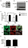
Figure 2. IGF-1 regulation by miR-1 on skeletal muscle C2C12 cells
(A) Western blot analysis for the C2C12 differentiation markers α-MHC and Troponin T. (B) mir-1 expression in C2C12 cells after differentiation into myotubes. Cells were switched to differentiation medium (DM) at day 0. (C) Luciferase reporter assay (mean ± S.E., minimun of 3 experiments per group) on C2C12 cells cultured in GM and DM with a luciferase reporter gene linked to wild-type (wt) or mutated (mt) 3′UTR of IGF-1. *P<0.05, versus controls (wt 3′UTR in GM or mt 3′UTR in DM). (D) Immunofluorescence staining for IGF-1 and F-actin of C2C12 cells cultured in growth medium (GM) and DM for 2 days. (E) qPCR for IGF-1 on C2C12 cells cultured in GM and DM. GAPDH was used as internal control.
Figure 3
Figure 3. Reciprocal regulation of miR-1 and IGF-1 expression in TAC and AKT overexpression models of cardiac hypertrophy
(A) Northern blot analysis and relative expression values of miR-1 in Sham-operated and transverse aortic arch-constricted (TAC) mice (representative results, mean ± S.E. of a minimum of n = 5 mice per group). (B) Western blot analysis for IGF-1 in left ventricles from Sham and TAC operated mice (representative results, mean ± S.E. of a minimum o f n = 5 mice per group). Band intensities were quantified using ImageJ software version 1.34 (http://rsb.info.nih.gov/ij/) and normalized to GAPDH. (C) miR-1 qRT-PCR on AKT and WT mice. (D) Western blot analysis for IGF-1 expression on AKT and WT mice (representative results, mean ± S.E. of a minimum of n = 5 mice per group). * P<0.05 versus controls (Sham or WT).
Figure 4
Figure 4. Regulation of miR-1 by the IGF-1 pathway
(A) miR-1 qRT-PCR on neonatal cardiomyocytes (nCMC) treated with 10 nM IGF-1 at different time points. NT: nCMC not treated. Sno202 RNA was utilized as internal control. (B) miR-1 qRT-PCR on differentiated C2C12 cells treated with various doses of IGF-1 for 2 days. NT: C2C12 not treated; DM NT: C2C12 differentiated not treated. DM: C2C12 differentiated. Sno202 RNA was utilized as internal control. (C) miR-1 qRT-PCR on nCMC infected with Ad-DA-AKT and Ad–DN-AKT at different multiciplicities of infection (m.o.i) (,Ad-Empty m.o.i 100;, Ad-Empty m.o.i 90 + Ad-AKT (DA or DN) m.o.i. 10;, Ad-Empty m.o.i 50 + Ad-AKT (DA or DN) m.o.i. 50;, Ad-AKT (DA or DN) m.o.i. 100). Sno202 RNA was utilized as internal control. *P<0.05 versus controls (nCMC NT, C2C12 DM NT and Ad-Empty). Data are expressed as the mean ± S.E. of 3 independent experiments.
Figure 5
Figure 5. Foxo3a regulates miR-1 expression
(A) The two potential Foxo3a binding sites on miR-1 promoter are indicated as BS1 and BS2. Both fragments of the miR-1 promoter were synthesized and linked to the luciferase (Luc) reporter gene. (B) 293T cells were infected with Ad-Empty or Ad-DA-foxo and Ad-DN-foxo at a m.o.i. of 50. 24 h after infection, cells were transfected with empty vector (pGL3.basic) or miR-1 promoter constructs, respectively. Firefly luciferase activities were normalized to Renilla luciferase activities. (C) nCMC were infected with Ad-DA-foxo and DN-foxo at different m.o.i (,Ad-Empty m.o.i 50;, Ad-Empty m.o.i 49 + Ad-foxo (DA or DN) m.o.i. 1;, Ad-Empty m.o.i 40 + Ad-foxo (DA or DN) m.o.i. 10;, Ad-foxo (DA or DN) m.o.i. 50). Cells were harvested 48 h after infection and analyzed for miR-1 levels by qRT-PCR. Sno202 RNA was utilized as internal control. (D) Western blot on C2C12 (DM1 and 3: differentiation medium at day 1 and 3) (Left Panel) and nCMC infected with Ad-miR-1 (m.o.i 100) (Right Panel) for Total AKT, P-AKT, Total Foxo3a and P-Foxo3a. Band intensities were quantified (bottom of each panel) as described in figure 3. *P<0.05 versus controls (Ad-Empty, Ad-DN-foxo). Data are expressed as the mean ± S.E. of 3 independent experiments.
Figure 6
Figure 6. miR-1 expression in acromegalic patients
(A) Left ventricular biopsy (Panel A) from a patient with acromegalic cardiomyopathy showing remarkable cardiomyocyte hypertrophy compared to normal control (Panel B) (H&E staining, 250X magnification). (B) miR-1 qRT-PCR on heart biopsies from acromegaly patients compared to healthy donors. U6 RNA was utilized as internal control. (C) Pearson product moment linear correlation between the LV (left ventricular) mass index, MWT (maximal wall thickness) and the level of miR-1 in acromegaly patients. *P<0.05 versus control (Healthy donors). Data are expressed as the mean ± S.E. of 3 independent experiments.
Figure 7
Figure 7. Proposed model of the miR-1/IGF-1 regulatory loop

References

    1. Bartel DP. MicroRNAs: genomics, biogenesis, mechanism, and function. Cell. 2004;116:281–297. - PubMed
    1. Cheng AM, Byrom MW, Shelton J, Ford LP. Antisense inhibition of human miRNAs and indications for an involvement of miRNA in cell growth and apoptosis. Nucleic Acids Res. 2005;33:1290–1297. - PMC - PubMed
    1. Felli N, Fontana L, Pelosi E, Botta R, Bonci D, Facchiano F, Liuzzi F, Lulli V, Morsilli O, Santoro S, Valtieri M, Calin GA, Liu CG, Sorrentino A, Croce CM, Peschle C. MicroRNAs 221 and 222 inhibit normal erythropoiesis and erythroleukemic cell growth via kit receptor down-modulation. Proc Natl Acad Sci U S A. 2005;102:18081–18086. - PMC - PubMed
    1. Xu P, Guo M, Hay BA. MicroRNAs and the regulation of cell death. Trends Genet. 2004;20:617–624. - PubMed
    1. Krek A, Grun D, Poy MN, Wolf R, Rosenberg L, Epstein EJ, MacMenamin P, da Piedade I, Gunsalus KC, Stoffel M, Rajewsky N. Combinatorial microRNA target predictions. Nat Genet. 2005;37:495–500. - PubMed

Publication types