. 2010 Feb;59(2):358-74.
doi: 10.2337/db09-1159. Epub 2009 Nov 23.

Cytokines interleukin-1beta and tumor necrosis factor-alpha regulate different transcriptional and alternative splicing networks in primary beta-cells

Affiliations

Cytokines interleukin-1beta and tumor necrosis factor-alpha regulate different transcriptional and alternative splicing networks in primary beta-cells

Fernanda Ortis et al. Diabetes. 2010 Feb.

Abstract

Objective: Cytokines contribute to pancreatic beta-cell death in type 1 diabetes. This effect is mediated by complex gene networks that remain to be characterized. We presently utilized array analysis to define the global expression pattern of genes, including spliced variants, modified by the cytokines interleukin (IL)-1beta + interferon (IFN)-gamma and tumor necrosis factor (TNF)-alpha + IFN-gamma in primary rat beta-cells.

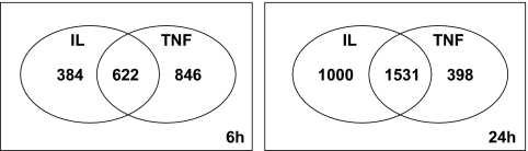
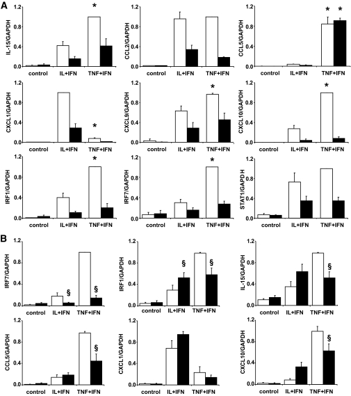
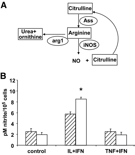
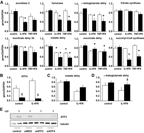
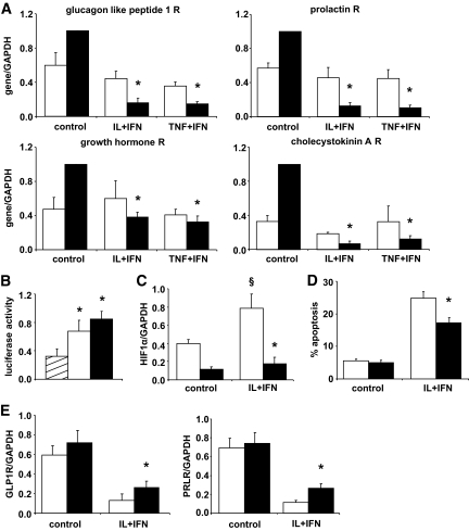
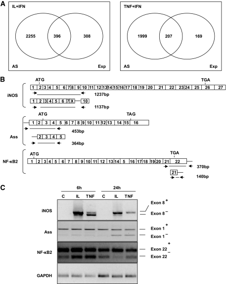
Research design and methods: Fluorescence-activated cell sorter-purified rat beta-cells were exposed to IL-1beta + IFN-gamma or TNF-alpha + IFN-gamma for 6 or 24 h, and global gene expression was analyzed by microarray. Key results were confirmed by RT-PCR, and small-interfering RNAs were used to investigate the mechanistic role of novel and relevant transcription factors identified by pathway analysis. RESULTS Nearly 16,000 transcripts were detected as present in beta-cells, with temporal differences in the number of genes modulated by IL-1beta + IFNgamma or TNF-alpha + IFN-gamma. These cytokine combinations induced differential expression of inflammatory response genes, which is related to differential induction of IFN regulatory factor-7. Both treatments decreased the expression of genes involved in the maintenance of beta-cell phenotype and growth/regeneration. Cytokines induced hypoxia-inducible factor-alpha, which in this context has a proapoptotic role. Cytokines also modified the expression of >20 genes involved in RNA splicing, and exon array analysis showed cytokine-induced changes in alternative splicing of >50% of the cytokine-modified genes.

Conclusions: The present study doubles the number of known genes expressed in primary beta-cells, modified or not by cytokines, and indicates the biological role for several novel cytokine-modified pathways in beta-cells. It also shows that cytokines modify alternative splicing in beta-cells, opening a new avenue of research for the field.


Figures

FIG. 1.
FIG. 1.
Effects of cytokine exposure on gene expression in FACS-purified rat β-cells. Ven diagram showing the number of β-cell genes with the expression modified by cytokines after exposure to IL-1β + IFN-γ (IL) or TNF-α + IFN-γ (TNF) for 6 and 24 h. The diagram shows genes modified by IL-1β + IFN-γ alone (left part of the figure), TNF-α + IFN-γ alone (right) or both (center). Results of three independent array experiments were analyzed. mRNA expression was considered as modified by cytokines when P < 0.02 and fold change ≥1.5 compared with control condition.
FIG. 2.
FIG. 2.
TNF-α and IL-1β differentially modulate expression of β-cell genes involved in the inflammatory response. Gene expression was analyzed by real-time RT-PCR. A: FACS-purified rat β-cells were exposed or not (control) to IL-1β + IFN-γ (IL+IFN) or to TNF-α + IFN-γ (TNF+IFN) for 6 h (□) or 24 h (■). B: FACS-purified rat β-cells were transfected with siRNA control (□) or siRNA against IRF-7 (■) and exposed or not (control) to IL-1β + IFN-γ (IL+IFN) or to TNF-α + IFN-γ (TNF+IFN) for 24 h. Results are means ± SE of three to six independent experiments. *P < 0.05 vs. IL+IFN at the same time point; §P < 0.05 vs. siControl at the same time point and treatment.
FIG. 3.
FIG. 3.
Differential usage of the NO synthesis pathway by IL-1β and TNF-α. A: Schematic view of the NO synthesis pathway. Elliptical shapes represent enzymes. B: Synthesis of NO by rat primary β-cells cultured in arginine-citrulline–free medium (▨) or in medium containing 1 mmol/l citrulline (□) and exposed to IL-1β + IFN-γ (IL+IFN) or TNF-α + IFN-γ (TNF+IFN) for 48 h. Results are mean of five independent experiments. *P < 0.05 vs. arginine-citrulline–free medium.
FIG. 4.
FIG. 4.
Cytokines decrease the expression of genes involved in the maintenance of a differentiated β-cell phenotype. Expression of genes related to β-cell function (A) or regulatory transcription factors (B) were analyzed by microarray (n = 3) in FACS-purified rat β-cells exposed to IL-1β + IFN-γ for 6 h (□) or 24 h (■) or to TNF-α + IFN-γ for 6 h (▨) or 24 h (vertical striped bars). Results are shown as fold change compared with control (no cytokine added), considered as one (line). C: confirmation by real-time RT-PCR of cytokine effects on the expression of PDX-1, MafA, and Isl1; □, 6 h; ■, 24 h. Results are means ± SE of three to four independent experiments. *P < 0.05; #P < 0.01; §P < 0.001 vs. control.
FIG. 5.
FIG. 5.
Cytokines inhibit expression of genes encoding Krebs cycle enzymes, which is partially dependent of ATF4 activation. A: Confirmation by real-time RT-PCR of microarray analysis data in rat purified β-cells untreated (control) or exposed to a combination of IL-1β + IFN-γ (IL+IFN) or TNF-α + IFN-γ (TNF+IFN) for 6 h (□) or 24 h (■). Results are means ± SE of four to five independent experiments. *P ≥ 0.05 vs. control. B and C: β-Cells were transfected with siControl (□) or siATF4 (■) and then left untreated or treated with IL-1β + IFN-γ (IL+IFN) for 24 h. B: Confirmation of ATF4 knockdown (KD) by real-time RT-PCR. C: Effects of ATF4 KD on the expression of malate dehydrogenase and α-ketoglutarase dehydrogenase. Results are mean of six independent experiments. §P < 0.05 cytokines + siATF4 vs. cytokines + siControl. Dehy = dehydrogenase. E: Western blot for ATF3 protein in cells transfected with siATF4 or siATF3. The figure is representative of four independent experiments.
FIG. 6.
FIG. 6.
Cytokines decrease expression of key hormone receptor genes partially via HIF-1α induction. Rat purified β-cells were left untreated (control) or exposed to a combination of IL-1β + IFN-γ (IL+IFN) or TNF-α + IFN-γ (TNF+IFN). A: Real-time RT-PCR to confirm microarray analysis of hormone receptors (R) expression after cytokine exposure for 6 h (□) or 24 h (■). Results are means ± SE of three to four experiments. *P ≤ 0.05 vs. control. B: Luciferase reporter assay of HIF-1α activation by cytokines. Cells were cotransfected with an HRE luciferase reporter gene and the internal control pRL-CMV, then left untreated (▨) or exposed to IL+IFN (□) or the positive control Cobalt chloride (CoCl2, ■) for 12 h. Results are normalized for Renilla luciferase activity and are means ± SE of five experiments. *P < 0.05 vs. untreated cells. C–E: HIF-1α knockdown by siRNA. Cells were transfected with siControl (□) or siHIF-1α (■) and then left untreated or treated with IL+IFN for 24 h. Results are means ± SE of four to six experiments. C: HIF-1α knockdown analyzed by real-time RT-PCR. *P ≤ 0.05 vs. siControl under the same treatment and §P ≤ 0.05 vs. control (not cytokine treated). D: Viability of cells after HIF-1α knockdown and 48 h cytokine exposure. *P ≤ 0.05 vs. siControl + cytokines. E: Expression of PRLR and GLP-1R after HIF-1α knockdown and 24-h cytokine exposure, measured by real-time RT-PCR. *P ≤ 0.05 vs. siControl + cytokines.
FIG. 7.
FIG. 7.
Cytokines induce alternative splicing in rat pancreatic β-cells. A: Ven diagram representing the number of β-cell genes that undergo alternative splicing (alternative splicing) and/or expression (Exp) changes after 24 h of cytokine treatment compared with control condition, as identified by exon array analysis (GeneChip Rat exon 1.0 ST Array). B: Schematic diagram of inducible iNOS, ASS, and NFκB subunit p100/p52 exon structures and of the PCR primers presently used to identify spliced forms. Start (ATG) and stop (TGA or TAG) codons are indicated in the figure. The arrows show the positions of the PCR primers, while the lines below indicate the size of the amplified region in the presence or absence of the respective exon analyzed. C: RT-PCR of rat primary β-cells exposed to control condition (C), IL-1β + IFN-γ (IL), or TNF-α + IFN-γ (TNF) for 6 or 24 h to amplify the regions of iNOS, ASS, and NF-κB indicated in B. GAPDH was amplified in parallel to control for the amount of cDNA loaded in each reaction. The figure is representative of three to five experiments.

Comment in

References

    1. Eizirik DL, Mandrup-Poulsen T: A choice of death-the signal-transduction of immune-mediated β-cell apoptosis. Diabetologia 2001;44:2115–2133 - PubMed
    1. Kaminitz A, Stein J, Yaniv I, Askenasy N: The vicious cycle of apoptotic β-cell death in type 1 diabetes. Immunol Cell Biol 2007;85:582–589 - PubMed
    1. Uno S, Imagawa A, Okita K, Sayama K, Moriwaki M, Iwahashi H, Yamagata K, Tamura S, Matsuzawa Y, Hanafusa T, Miyagawa J, Shimomura I: Macrophages and dendritic cells infiltrating islets with or without β-cells produce tumour necrosis factor-α in patients with recent-onset type 1 diabetes. Diabetologia 2007;50:596–601 - PubMed
    1. Mastrandrea L, Yu J, Behrens T, Buchlis J, Albini C, Fourtner S, Quattrin T: Etanercept treatment in children with new-onset type 1 diabetes: pilot randomized, placebo-controlled, double-blind study. Diabetes Care 2009;32:1244–1249 - PMC - PubMed
    1. Pickersgill LM, Mandrup-Poulsen TR: The anti-interleukin-1 in type 1 diabetes action trial-background and rationale. Diabetes Metab Res Rev 2009;25:321–324 - PubMed

Publication types

MeSH terms