. 2009 Dec;136(24):4155-64.
doi: 10.1242/dev.034355.

N-cadherin acts in concert with Slit1-Robo2 signaling in regulating aggregation of placode-derived cranial sensory neurons

Affiliations

N-cadherin acts in concert with Slit1-Robo2 signaling in regulating aggregation of placode-derived cranial sensory neurons

Celia E Shiau et al. Development. 2009 Dec.

Abstract

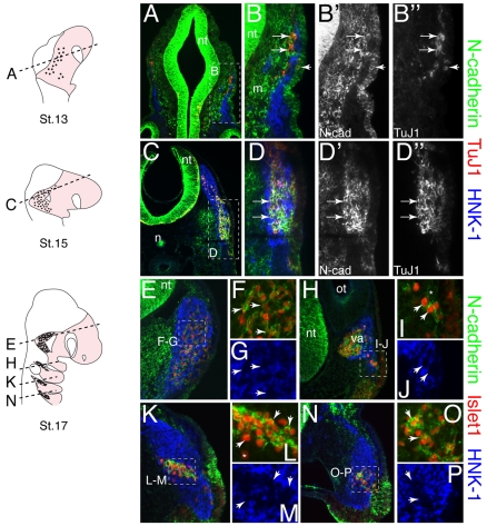
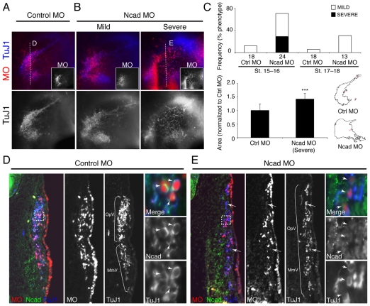
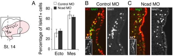
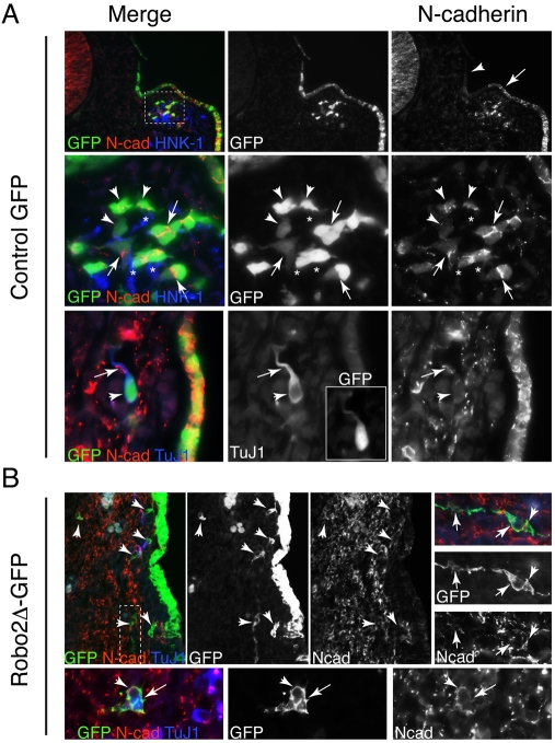
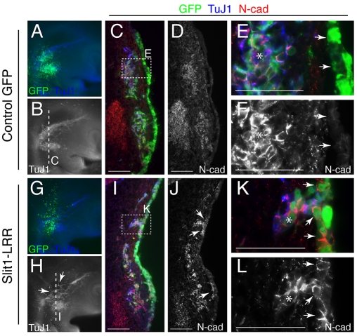
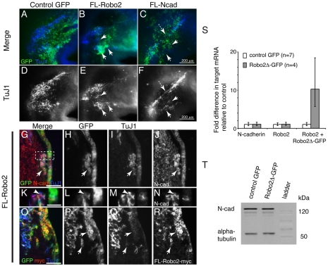
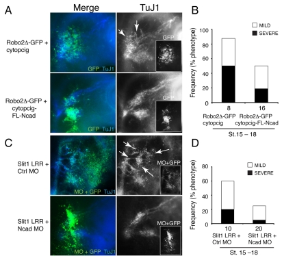
Vertebrate cranial sensory ganglia have a dual origin from the neural crest and ectodermal placodes. In the largest of these, the trigeminal ganglion, Slit1-Robo2 signaling is essential for proper ganglion assembly. Here, we demonstrate a crucial role for the cell adhesion molecule N-cadherin and its interaction with Slit1-Robo2 during gangliogenesis in vivo. A common feature of chick trigeminal and epibranchial ganglia is the expression of N-cadherin and Robo2 on placodal neurons and Slit1 on neural crest cells. Interestingly, N-cadherin localizes to intercellular adherens junctions between placodal neurons during ganglion assembly. Depletion of N-cadherin causes loss of proper ganglion coalescence, similar to that observed after loss of Robo2, suggesting that the two pathways might intersect. Consistent with this possibility, blocking or augmenting Slit-Robo signaling modulates N-cadherin protein expression on the placodal cell surface concomitant with alteration in placodal adhesion. Lack of an apparent change in total N-cadherin mRNA or protein levels suggests post-translational regulation. Co-expression of N-cadherin with dominant-negative Robo abrogates the Robo2 loss-of-function phenotype of dispersed ganglia, whereas loss of N-cadherin reverses the aberrant aggregation induced by increased Slit-Robo expression. Our study suggests a novel mechanism whereby N-cadherin acts in concert with Slit-Robo signaling in mediating the placodal cell adhesion required for proper gangliogenesis.


Figures

Fig. 1.
Fig. 1.
Expression of chick N-cadherin protein on trigeminal and epibranchial ectodermal and ingressing placode-derived neurons. Schematics on left indicate the levels of the sections (dashed lines), the neural crest region (pink) and placodes (black dots). Placode-derived neurons are marked by β-neurotubulin (TuJ1) or Islet1, and neural crest cells by HNK-1. (A) Stage 13 showing ingressing trigeminal placodes. (B-B″) Higher magnification of boxed region in A. Ingressed placodal cells (arrows) and ectodermal placodes (arrowhead) co-express TuJ1 and N-cadherin. (C-D″) Stage 15 in the early forming trigeminal ganglion. N-cadherin expression overlaps with the placodal marker TuJ1 (arrows), but not with HNK-1, at all stages. (E-P) Stage 17 in the condensing (E-G) trigeminal, (H-J) facial (or geniculate), (K-M) glossopharyngeal (or petrosal), and (N-P) vagal (or nodose) ganglionic regions. Few Islet1-negative cells express N-cadherin at stage 17 (I, star). Islet1+ cells co-express N-cadherin (arrowheads), with some exceptions of individual placodal cells (L, star). HNK-1+ neural crest cells do not express N-cadherin at all cranial axial levels. N-cad, N-cadherin; nt, neural tube; m, mesoderm; n, notochord; ot, otic vesicle; va, vestibuloacoustic ganglion.
Fig. 2.
Fig. 2.
N-cadherin knockdown blocks proper trigeminal placodal aggregation. (A) Control MO trigeminal ganglion. (B) Ncad MO showing ‘mild’ and ‘severe’ phenotypes at stages 15-16. (C) (Above) A high percentage of ganglionic defects in Ncad MO embryos at stages 15-16 and a lower percentage at stages 17-18, with little effect in controls. The number ganglia analyzed (n) is shown beneath each bar. (Below) Significantly more dispersed trigeminal placodal ganglia in severely affected Ncad MO embryos (1.42±0.21) relative to controls (1.0±0.24) (***P=0.0003, two-tailed Student's t-test) using ImageJ area analysis. Error bars, s.d. To the right are shown representative outlines of calculated placodal areas. (D,E) Frontal plane sections through the same embryos as in A and B at the level indicated by the dashed lines. Placodal neurons in the Ncad MO ganglion were markedly more scattered. Panels showing MO alone indicate the area of transfection, and those showing TuJ1 alone indicate the distribution of placodes. The boxed region is shown at higher magnification to the right; arrows indicate N-cadherin reduction in Ncad MO but not control MO placodes. (E) A few scattered placode-derived neurons do not have Ncad MO (arrows). OpV and MmV lobes are not distinct in the Ncad MO embryo, as compared with the control (brackets). Ctrl, control; Ncad, N-cadherin; MO, morpholino; OpV, ophthalmic; MmV, maxillomandibular.
Fig. 3.
Fig. 3.
Morpholino-mediated knockdown of N-cadherin does not block placodal ingression. To the left is a schematic of a stage 14 chick embryo showing the region of analysis (boxed) and the level of sections in B and C (dashed line). (A) Bar chart showing that the percentages of Islet1+ placodal cells associated with ectoderm and in the mesenchyme were not significantly different between control MO (n=4) and Ncad MO (n=3) embryos (P=0.43, two-tailed Student's t-test). Error bars, s.d. (B,C) Left, overlay showing MO transfection (red and inset) and Islet1 (green). Right, single-channel image of Islet1. Compared with control MO embryos (B), placodal cells in the Ncad MO embryos (C) appear more scattered early on at stage 14. Ecto, ectoderm; Mes, mesenchyme.
Fig. 4.
Fig. 4.
Robo2 inhibition blocks formation of N-cadherin-localized tight adherens junctions. (A) (Top row) Cross-section through a stage 13 control GFP chick embryo showing N-cadherin protein expression in the dorsomedial and ventral ectoderm (arrow) where placodal neurons arise, and not in the most dorsal ectoderm (arrowhead). (Middle row) Higher magnification of the boxed region showing N-cadherin localized at sites of placodal cell-cell contacts (GFP+, arrows) and punctate expression on individual placodal neurons (GFP+, arrowheads). HNK-1+ neural crest cells (blue, stars) associate closely with placodal cells. (Bottom row) N-cadherin is largely downregulated on the cell body of individual placodal neurons (GFP+/TuJ1+), although weak punctate spots remain (arrowhead), and there is prominent expression at the contact site between the neuronal process and another cell (arrow). (B) (Top row) Robo2-blocked placodal neurons (GFP+/TuJ1+) are more dissociated and do not have high N-cadherin expression at the cell boundaries (arrowheads). The boxed region is shown (rotated 90 degrees) at higher magnification to the right, showing ingressed Robo2Δ-GFP placodal cells expressing little or no N-cadherin (arrows) and punctate N-cadherin at the cell junction (arrowhead). (Bottom row) Two associating placodal cells blocked by Robo2Δ-GFP do not form N-cadherin-localized adherens junctions (arrow) but still express some N-cadherin on the peripheral cell surface (arrowhead).
Fig. 5.
Fig. 5.
Ectopic Slit1 expression in the placodal ectoderm leads to increased N-cadherin expression at sites of aberrant placodal clusters. (A,B) Control GFP chick embryo showing the trigeminal placodal ganglion (GFP+/TuJ1+). (C) Frontal plane section through the two trigeminal lobes as indicated in B. (D) N-cadherin protein expression in the same section as in C. (E,F) Higher magnification of boxed region in C showing downregulation of N-cadherin in the surface ectoderm (arrows) but strong expression in the ingressed placodal cells (asterisk). (G,H) Slit1-LRR-transfected ganglion showing aberrant aggregation of placodal cells (arrows, GFP+/TuJ1+). (I,J) Frontal plane section as indicated in H. (K,L) Higher magnification of boxed region in I; N-cadherin is abnormally upregulated in the surface ectoderm (arrows); N-cadherin is also highly expressed in the ingressed placodal cluster (asterisk). Scale bars: 100 μm in C,D,I,J; 50 μm in E,F,K,L.
Fig. 6.
Fig. 6.
Overexpression of either Robo2 or N-cadherin leads to abnormal aggregation and increased N-cadherin at contact sites, but total N-cadherin mRNA and protein levels are not altered by blocking Robo2. (A-F) Control GFP (A,D), full-length Robo2 (FL-Robo2) (B,E), and full-length N-cadherin (FL-Ncad) (C,F) chick embryos at stage 16. Overexpression of Robo2 (B,E) or N-cadherin (C,F) causes abnormal aggregation in the ganglionic anlage (arrows). N-cadherin overexpression also causes an apparent loss of placodal neurons (dashed arrow). (G-J) Sections through the FL-Robo2 embryo showing high N-cadherin expression (arrow) on aberrant placodal aggregates (TuJ1+/GFP+) near the ectoderm. (K-N) Higher magnification of the boxed area in G, showing high N-cadherin expression at placodal cell junctions (arrowhead). (O-R) Ectopic aggregates near the ectoderm and in the mesenchyme (arrows) express the FL-Robo2-myc protein near the coalescing ganglion (star). (S) There is no statistical difference in N-cadherin or endogenous Robo2 transcript levels between control (n=7) and Robo2-inhibited (n=4) embryos as assessed by qPCR. As a positive control, a significant (~10-fold on average) increase in the exogenous Robo2Δ-GFP transcript level was detected relative to the control. Error bars, s.d. For supporting details, see Fig. S4 in the supplementary material. (T) A representative western blot showing no detectable difference in the total N-cadherin protein level between Robo2Δ-GFP- and GFP-transfected stage 17 head tissues that encompass the trigeminal ganglia. Per lane, 12.5 μg sample was loaded, with α-tubulin as loading control. Scale bars: 50 μm in G-J,O-R; 20 μm in K-N.
Fig. 7.
Fig. 7.
Modulating N-cadherin expression reverses the effects of Robo2 inhibition and Slit1 overexpression. (A) (Top) Co-electroporation of Robo2Δ-GFP and empty vector (cytopcig) leads to severely disorganized and dispersed ganglia (arrows). (Bottom) Co-expression of Robo2Δ-GFP with full-length N-cadherin (cytopcig-FL-Ncad) suppresses the severe effects of blocking Robo2 function, giving rise to normally coalesced ganglia. Grayscale images show single channel of the placodal neuronal marker TuJ1 and of the transfection reporter GFP in the inset. (B) Co-expression of FL-Ncad and Robo2Δ-GFP significantly reduces the severe effects of Robo2Δ-GFP by 31% and overall by ~40%. Embryos scored as ‘mild’ have misshapen ganglia with aggregation defects in regions of the ganglia; those scored as ‘severe’ have widespread dispersed neurons and markedly abnormal ganglia in at least one lobe. (C) (Top) Co-electroporation of Slit1-LRR and control MO in the trigeminal placodal ectoderm leads to severely abnormal patterns of ganglion aggregation (arrows). (Bottom) Co-expression of Slit1-LRR with Ncad MO significantly decreases ganglion coalescence defects. Green, MO tagged with 3′ fluorescein and GFP; blue, TuJ1. (D) Co-expression of Slit1-LRR and Ncad MO significantly reduces the effects of Slit1 overexpression, overall by ~35%. Embryos scored as ‘mild’ have misshapen ganglia with aggregation defects; those scored as ‘severe’ have dramatic defects in condensation, including ectopic aggregates and/or axonal disorganization in at least one lobe. The number ganglia analyzed (n) is shown beneath each bar.

Similar articles

Cited by

References

    1. Akitaya T., Bronner-Fraser M. (1992). Expression of cell adhesion molecules during initiation and cessation of neural crest cell migration. Dev. Dyn. 194, 12-20 - PubMed
    1. Baker C. V. (2005). Neural crest and cranial ectodermal placodes. In Developmental Neurobiology (ed. Jacobson M. S. R. a. M.), pp. 67-127 New York: Kluwer Academic/Plenum Publishers;
    1. Battye R., Stevens A., Perry R. L., Jacobs J. R. (2001). Repellent signaling by Slit requires the leucine-rich repeats. J. Neurosci. 21, 4290-4298 - PMC - PubMed
    1. Begbie J., Graham A. (2001). Integration between the epibranchial placodes and the hindbrain. Science 294, 595-598 - PubMed
    1. Braga V. M. (1999). Small GTPases and regulation of cadherin dependent cell-cell adhesion. Mol. Pathol. 52, 197-202 - PMC - PubMed

Publication types

MeSH terms