. 2009 Dec 21;206(13):3031-46.
doi: 10.1084/jem.20091892. Epub 2009 Nov 23.

Both XPD alleles contribute to the phenotype of compound heterozygote xeroderma pigmentosum patients

Affiliations

Both XPD alleles contribute to the phenotype of compound heterozygote xeroderma pigmentosum patients

Takahiro Ueda et al. J Exp Med. .

Abstract

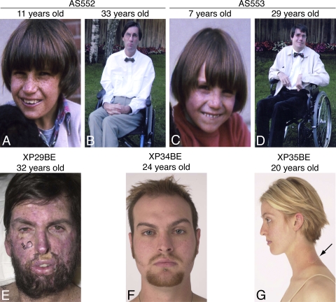
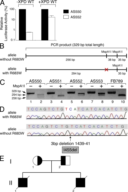
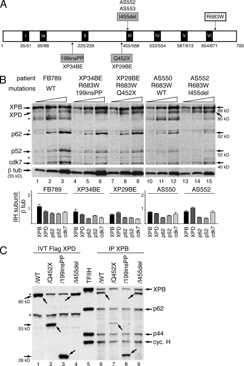
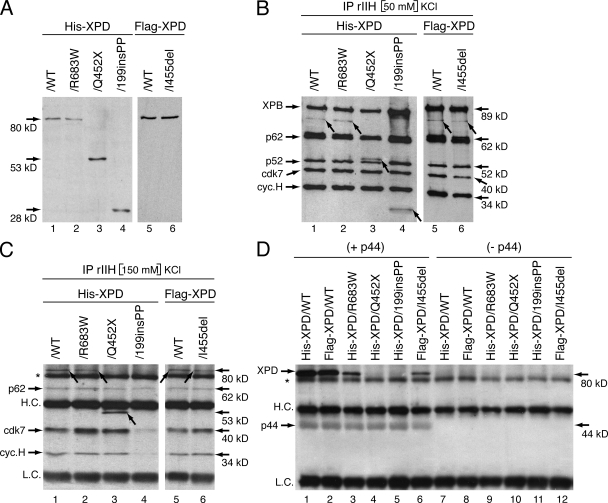
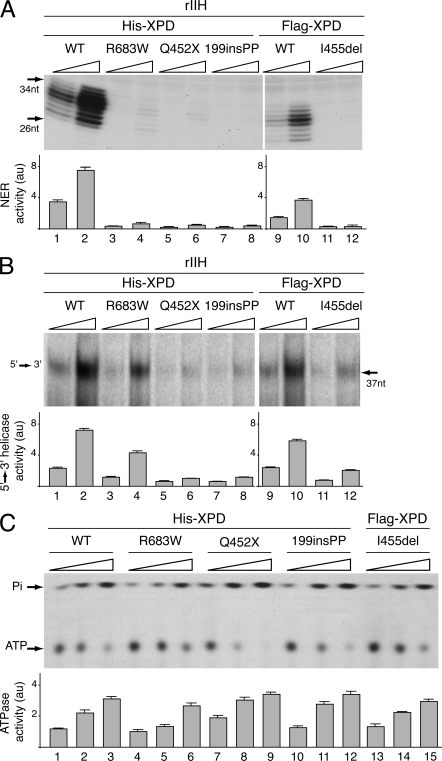
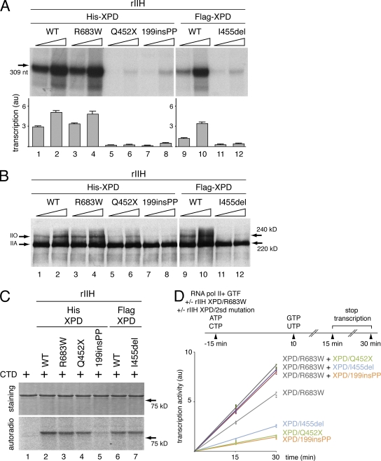
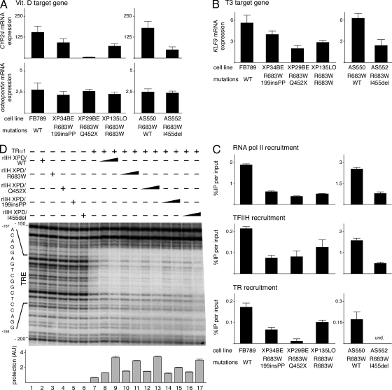
Mutations in the XPD subunit of the DNA repair/transcription factor TFIIH result in the rare recessive genetic disorder xeroderma pigmentosum (XP). Many XP patients are compound heterozygotes with a "causative" XPD point mutation R683W and different second mutant alleles, considered "null alleles." However, there is marked clinical heterogeneity (including presence or absence of skin cancers or neurological degeneration) in these XPD/R683W patients, thus suggesting a contribution of the second allele. Here, we report XP patients carrying XPD/R683W and a second XPD allele either XPD/Q452X, /I455del, or /199insPP. We performed a systematic study of the effect of these XPD mutations on several enzymatic functions of TFIIH and found that each mutation exhibited unique biochemical properties. Although all the mutations inhibited the nucleotide excision repair (NER) by disturbing the XPD helicase function, each of them disrupted specific molecular steps during transcription: XPD/Q452X hindered the transactivation process, XPD/I455del disturbed RNA polymerase II phosphorylation, and XPD/199insPP inhibited kinase activity of the cdk7 subunit of TFIIH. The broad range and severity of clinical features in XP patients arise from a broad set of deficiencies in NER and transcription that result from the combination of mutations found on both XPD alleles.


Figures

Figure 1.
Figure 1.
XP patients studied. (A and B) Patient AS552. (A) At 11 yr of age he had marked freckle-like hyperpigmentation in sun exposed portions of his face and neck. (B) By age 33 yr he used a wheelchair because of his inability to walk. He died at age 39 yr of progressive neurological degeneration. (C and D) Patient AS553, brother of AS552. (C) At 7 yr of age he had marked freckle-like hyperpigmentation in sun exposed portions of his face. (D) By 29 yr of age he used a wheelchair because of his inability to walk. He had posturing of his hands, a feature of neurodegeneration. He died at age 35 yr of progressive neurological degeneration. (E) Patient XP29BE at 32 yr of age. He has marked freckle-like hyperpigmentation in sun exposed portions of his face and neck. His face shows extensive scarring and grafting from numerous surgical procedures for removal of skin cancers. The circled lesions on his face and shoulder were being followed photographically for signs of skin cancer. His conjunctiva show telangiectasia. He died at age 37 yr of progressive neurological degeneration. (F) Patient XP34BE. He was well protected from sun exposure and at age 24 yr he had minimal freckle-like hyperpigmentation on his face and neck and no skin cancers. By age 31 yr he had no evidence of neurological degeneration. (G) Patient XP35BE, younger sister of patient XP34BE. She was well protected from sun exposure and at age 20 yr she had minimal freckle-like hyperpigmentation on her face and neck and no skin cancers. Brief sun exposure of an unprotected portion of her neck resulted in an acute sunburn followed by hyperpigmentation, scaling and peeling of the skin (arrow). By age 27 yr she had no evidence of neurological degeneration.
Figure 2.
Figure 2.
Identification of the XPD mutations in the AS550 and AS552 patients. (A) Host cell reactivation in AS550 (filled columns) and AS552 (open columns). Fibroblasts were transfected with the pCMV-luc (500 ng) previously exposed to 1,000 J/cm2 of UVC-light (254 nm), pCH110 (100 ng, encoding the β-galactosidase which was used to normalize transfection efficiencies), and either empty pcDNA (10 ng; left pair of columns) or pcDNA XPD WT (10 ng, XPD WT; right pair of columns). Host cell reactivation was measured as the relative luciferase activity caused by the repair of the UV-damaged luciferase gene. The values of three independent experiments are presented as percentages, 100% being the level of luciferase activity obtained in AS550 cells. (B) Scheme representing the PCR products obtained from XPD allele with or without the point mutation R683W, which corresponds to a C-to-T substitution at the first base of exon 22 in the gene. This mutation abolishes the restriction site for the endonuclease MspA1l. (C) PCR products were obtained from genomic DNA of AS550, AS551, AS552, AS553, and FB789 (used as control) fibroblasts. Restriction fragment length polymorphism assay was performed from the PCR products digested by MspA1l. (D) DNA sequencing of the PCR product obtained from cDNA of AS552 cells. The XPD allele without the R683W point mutation has a deletion of three bases (TCA) at position 1439–41, giving rise to an amino acid del455 in-frame (I455del). (E) Genetic pedigree of the family of the patients AS552 and AS553. The mother AS550 (I-1) carries the XPD mutation that corresponds to an amino acid substitution at position R683W. The father AS551 (I-2) carries a deletion of three bases (TCA) at position 1439–41 that makes amino acid del455 in-frame (I455del). The deceased patients AS552 (II-1) and his brother AS553 (II-2) had compound heterozygous mutations that corresponded to the substitution at R683W (maternal) and the deletion I455del (paternal), resulting in the development of XP with skin cancer and neurological degeneration.
Figure 3.
Figure 3.
TFIIH complex into XPD fibroblasts. (A) The diagram represents the 760-aa XPD protein with the 7 (I–VI) helicase motifs. Amino acid changes resulting from mutations found in the XPD/R683W patients who are compound heterozygotes are depicted. (B) Western blot analyses of TFIIH subunits (XPB, XPD, p62, p52, and cdk7) in increasing amounts of whole-cell extracts isolated from fibroblasts of FB789, XP34BE, XP29BE, AS550, and AS552 patients. * indicate nonspecific bands. β-Tubulin (β tub) was used as an internal control. The results are representative of three independent experiments. Diagram represents the ratio between each TFIIH subunit and β tub (arbitrary units). (C) Western blotting analysis of Ab-XPB ChIP (IP XPB) samples from chromatin extracts isolated from HD2 cells transfected with expression vectors encoding either Flag-XPD/WT (∼80 kD), /Q452X (∼53 kD), /199insPP (∼28 kD), or /I455del (∼80 kD). The immunoprecipitated fractions were resolved on SDS-PAGE followed by immunoblotting using antibodies raised against XPB, p62, p44, cyclin H and the Flag-tag. In vitro synthesized Flag-XPD mutated proteins (IVT Flag XPD, lanes 1 to 4) and highly purified TFIIH from HeLa cells (lane 5) were used as references. Arrows indicate the different forms of XPD. * indicate nonspecific bands. The results are representative of three independent experiments.
Figure 4.
Figure 4.
XPD mutations disrupt TFIIH integrity. (A) Production in SF9 insect cells of the recombinant mutated forms of XPD tagged with either the His-tag (His-XPD/WT, /R683W, Q452X, and 199insPP) or the Flag-tag (Flag-XPD/WT and /I455del). Whole-cell extracts were resolved by SDS-PAGE and blotted with antibodies raised against either the His- or the Flag-tag. Arrows indicate the theoretical molecular weight of each XPD mutated form. The results are representative of four independent experiments. (B) Purification of the recombinant TFIIH (rIIH). Insect cells were infected with baculoviruses overexpressing the subunits of TFIIH including either WT or mutated XPD, and complexes were immunoprecipitated by using antibody (Ab) directed toward the p44 subunit of the core TFIIH in low salt conditions (50mM KCl). After elution with a synthetic peptide recognized by Ab-p44, equal amounts of purified rIIHs were then resolved by SDS-PAGE and blotted with antibodies against XPB, p62, p52, cdk7 and cyclin H subunits of TFIIH. His-XPD/WT, /R683W, /Q452X, /199insPP, and Flag-XPD/WT, /I455del were visualized with antibodies raised against either the His- or the Flag-tag, respectively. Arrows indicate the different forms of XPD. The results are representative of five independent experiments. (C) Immunoprecipitation with Ab-p44 of the various rIIH in a higher salt condition (150 mM KCl). rIIHs immunoprecipitated with Ab-p44 cross-linked on agarose beads were boiled, resolved by SDS-PAGE, and blotted with antibodies against p62, cdk7, cyclin H, and either the His- or the Flag-tag. Arrows show the different forms of XPD. HC., heavy chain of Ab-p44; LC, light chain of Ab-p44; * indicates a nonspecific band. The results are representative of three independent experiments. (D) Infected Sf9 cell lysates containing either WT or mutated XPD as indicated at the top of the panel, were incubated with (+ p44) or without (− p44) WT p44, immunoprecipitated to agarose beads and further washed with 350 mM KCl. The immunoprecipitated fractions were then resolved on SDS-PAGE, followed by immunoblotting using Ab-p44 and either anti-His or anti-Flag antibodies. * indicates a nonspecific band. The results are representative of three independent experiments.
Figure 5.
Figure 5.
DNA Repair Activities of the rIIHs. (A) Equivalent increasing amounts of rIIHs (adjusted according to Western blotting) were added to an incision/excision assay using recombinant NER factors. The area of the gel containing the excision products is shown (the complete gel is shown in Fig. S1 A). The excised oligonucleotides signals were quantified and plotted in arbitrary units (au). The results are representative of three independent experiments. (B) Helicase activity of the WT and mutated forms of XPD. Equivalent increasing amounts of the various immunoprecipitated rIIH were tested for their 5′–3′ XPD helicase activity. The density of radiolabeled oligo displaced by the XPD helicase activity was measured and plotted in arbitrary units (au). The results are representative of three independent experiments. The complete gel is shown in Fig. S1 B. (C) Equal amounts of immunoprecipitated XPD were tested in an ATPase assay for 120 min. Nonhydrolyzed ATP as well as Pi are indicated. The ATP hydrolysis was quantified and plotted in arbitrary units (au). The results are representative of three independent experiments.
Figure 6.
Figure 6.
Transcription activity of the rIIHs. (A) Basal transcription activity of the rIIHs. The purified rIIHs were added to an in vitro reconstituted transcription system lacking TFIIH. The length of the corresponding transcript is indicated on the left side. The transcription activity of all variants was assessed using increasing amounts of rIIHs for 30 min. The signals were quantified and plotted in arbitrary units (au). The results are representative of three independent experiments. The complete gel is shown in Fig. S2 A. (B) Phosphorylation of the RNA polymerase II during in vitro reconstituted transcription assays. The RNA pol II kinase activity of low salt immunopurifed rIIHs was analyzed in an in vitro assay containing all the basal transcription factors and the AdMLP. Arrows indicate hypo (IIA) and hyper (IIO) phosphorylated forms of RNA pol II. The results are representative of two independent experiments. The complete gel is shown in Fig. S2 B. (C) In vitro phosphorylation of the GST-CTD fusion protein was performed with equal amounts of rIIHs in the presence of 0.14 µM [γ-32P] ATP. Coomassie blue–stained gel (staining; top) and autoradiography (autoradio; bottom) of the incubated fractions are shown. The results are representative of three independent experiments. The complete gels are shown in Fig. S2 C. (D) In vitro reconstituted transcription assays were performed following the protocol scheme. rIIH XPD/R683W, rIIH XPD/Q452X, /199insPP, or /I455del were preincubated either alone or in combination, in the presence of RNA pol II, the general transcription factors (GTF), the AdMLP template, ATP, and CTP. 15 min later, the transcription process was initiated by addition of GTP and UTP. The reactions were performed for 15 and 30 min. The signals of three independent experiments were then quantified and plotted in arbitrary units (au).
Figure 7.
Figure 7.
Incidence of XPD mutations during the transactivation mediated by nuclear receptors. (A) Expression of the VDR target genes CYP24 and osteopontin in WT (FB789, AS550) and XPD mutated (XP34BE, XP29BE, XP135LO, AS552) fibroblasts after treatment during 8h with vitamin D (vit. D, 100 nM). The values were normalized relative to 18S RNA expression. The results of three independent experiments are presented as n-fold induction relative to nontreated cells. (B) Expression of the TR target gene KLF9 in FB789, XP34BE, XP29BE, XP135LO, AS550, and AS552 fibroblasts after treatment during 24 h with T3 (10 nM). The values were normalized relative to 18S RNA expression. The results of three independent experiments are presented as n-fold induction relative to nontreated cells. (C) Recruitment of RNA pol II, TFIIH, and TR on the KLF9 promoter. After T3 treatment, the recruitment of RNA pol II and TFIIH (via its p44 subunit) was analyzed by ChIP assays on the KLF9 proximal promoter. The TR recruitment was studied on a TRE located at −2.9 kb in the KLF9 promoter. The results of three independent experiments are presented as percentage of DNA immunoprecipitated relative to the input; und, undetectable. (D) DNase1 footprint analysis on the TRE located into the TR target gene mbp. The MBP fragment (−256/+21) was labeled at the 5′ end and incubated with TRα1 and increasing amounts of rIIH XPD/WT (lanes 8–9), /R683W (lanes 10 and 11), /Q452X (lanes 12 and 13), /199insPP (lanes 14 and 15), and /I455del (lanes 16 and 17). Cis-element for the TRs (from nt-184 to nt-167) is positioned. Autoradiography of the gel is shown from nt-200 to nt-150. Graph depicts the protection level on the MBP-TRE (see Materials and methods). The complete gel is shown in Fig. S3 A.

Similar articles

Cited by

References

    1. Akeno N., Saikatsu S., Kawane T., Horiuchi N. 1997. Mouse vitamin D-24-hydroxylase: molecular cloning, tissue distribution, and transcriptional regulation by 1alpha,25-dihydroxyvitamin D3. Endocrinology. 138:2233–2240 10.1210/en.138.6.2233 - DOI - PubMed
    1. Akoulitchev S., Mäkelä T.P., Weinberg R.A., Reinberg D. 1995. Requirement for TFIIH kinase activity in transcription by RNA polymerase II. Nature. 377:557–560 10.1038/377557a0 - DOI - PubMed
    1. Andressoo J.O., Jans J., de Wit J., Coin F., Hoogstraten D., de Ven M.V., Toussaint W., Huijmans J., Thio H.B., van Leeuwen W.J., et al. 2006. Rescue of progeria in trichothiodystrophy by homozygous lethal Xpd alleles. PLoS Biol. 4:e322 10.1371/journal.pbio.0040322 - DOI - PMC - PubMed
    1. Araújo S.J., Tirode F., Coin F., Pospiech H., Syväoja J.E., Stucki M., Hübscher U., Egly J.M., Wood R.D. 2000. Nucleotide excision repair of DNA with recombinant human proteins: definition of the minimal set of factors, active forms of TFIIH, and modulation by CAK. Genes Dev. 14:349–359 - PMC - PubMed
    1. Botta E., Nardo T., Lehmann A.R., Egly J.M., Pedrini A.M., Stefanini M. 2002. Reduced level of the repair/transcription factor TFIIH in trichothiodystrophy. Hum. Mol. Genet. 11:2919–2928 10.1093/hmg/11.23.2919 - DOI - PubMed

Publication types

Substances