. 2009 Dec 21;206(13):2907-14.
doi: 10.1084/jem.20091225. Epub 2009 Nov 23.

Germinal center reutilization by newly activated B cells

Affiliations

Germinal center reutilization by newly activated B cells

Tanja A Schwickert et al. J Exp Med. .

Abstract

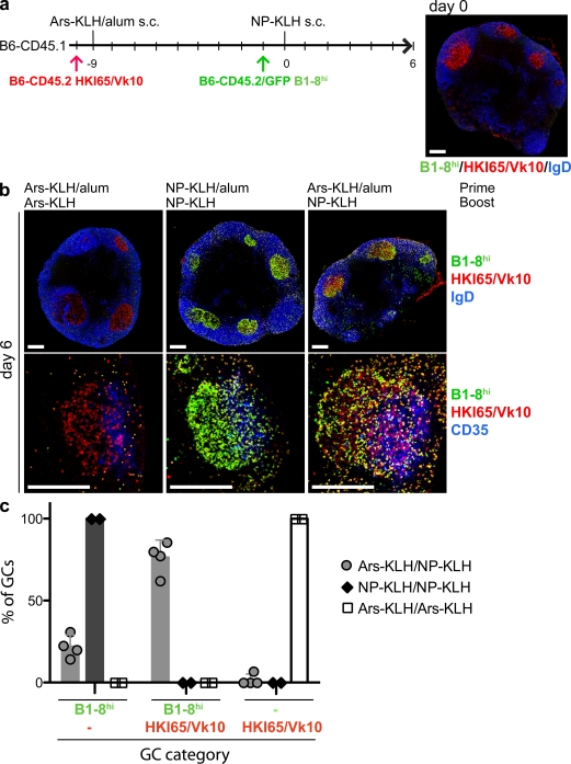
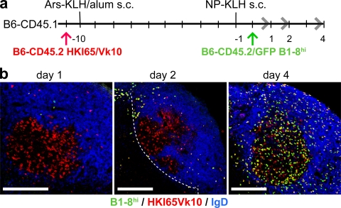
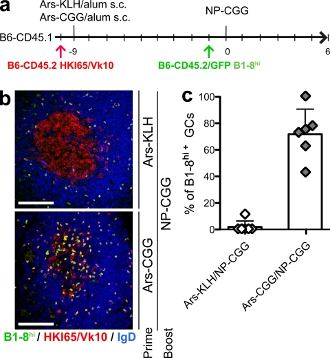
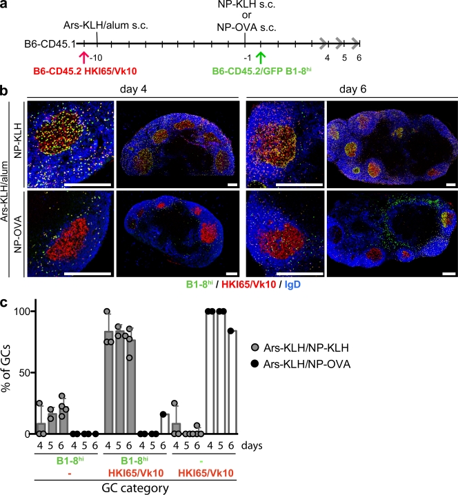
Germinal centers (GCs) are specialized structures in which B lymphocytes undergo clonal expansion, class switch recombination, somatic hypermutation, and affinity maturation. Although these structures were previously thought to contain a limited number of isolated B cell clones, recent in vivo imaging studies revealed that they are in fact dynamic and appear to be open to their environment. We demonstrate that B cells can colonize heterologous GCs. Invasion of primary GCs after subsequent immunization is most efficient when T cell help is shared by the two immune responses; however, it also occurs when the immune responses are entirely unrelated. We conclude that GCs are dynamic anatomical structures that can be reutilized by newly activated B cells during immune responses.


Figures

Figure 1.
Figure 1.
B cell invasion of established GCs. (a) Time line of immunization and B cell transfer. Ars-specific B cells (B6-CD45.2 HKI65/Vk10) were transferred 1 d before primary immunization (day −10) and NP-specific B cells (B6-CD45.2/GFP B1-8hi) were transferred 1 d before boosting (day −1) into wild-type (B6-CD45.1) mice. Ars-specific GCs were detected in histological sections, 9 d after priming (day 0; right: B6-CD45.2/GFP B1-8hi [green and red], B6-CD45.2 HKI65/Vk10 [red], IgD [blue]). (b) Representative histological sections of lymph nodes harvested 6 d after NP-KLH boost. Both transferred B cell populations (HKI65/Vk10 and B1-8hi) are membrane stained by CD45.2 (red), whereas B1-8hi B cells are additionally GFP+ (green; B1-8hi appear yellow because of GFP/CD45.2 double staining). To visualize GCs within secondary follicles, mantle zone B cells were stained by IgD (blue, top) or FDCs were stained by CD35 (blue, bottom). The top row shows whole cross sections of popliteal lymph nodes, and the bottom row shows more magnified views of GCs. Priming and boosting antigens are indicated at the top. Bars, 200 µm. (c) Statistical analysis of data shown in b. GCs were categorized in three groups: GCs with only NP-specific B cells, with both NP- and Ars-specific B cells, or with only Ars-specific B cells. Each data point represents the percentage of GCs in one of the categories for each mouse. Error bars show the standard deviation. Two independent experiments were performed with a total of four mice (two mice per control).
Figure 2.
Figure 2.
Kinetics of GC invasion. (a) Time line of immunization and B cell transfer. Ars-specific B cells (B6-CD45.2 HKI65/Vk10) were transferred into wild-type (B6-CD45.1) mice. 11 d later (day 0), NP-specific B cells (B6-CD45.2/GFP B1-8hi) were transferred. Mice were immunized with Ars-KLH in alum 24 h after Ars-specific cells were transferred. 1 d before the transfer of NP-specific B cells, mice were boosted with NP-KLH. (b) Histological sections of lymph nodes harvested at 1, 2, and 4 d after NP-KLH boost stained with CD45.2 (red) and IgD (blue) to label all transferred B cells (HKI65/Vk10 and B1-8hi) and naive B cells, respectively. Transferred NP-specific B1-8hi B cells were additionally GFP+ (green). Representative images of secondary follicles from two independent experiments are displayed. The dashed line indicates the T/B cell interface. Bars, 200 µm.
Figure 3.
Figure 3.
Invasion of heterogeneous GCs in the presence and absence of preexisting T cell help. (a) Time line of immunization and B cell transfer. Ars-specific B cells (B6-CD45.2 HKI65/Vk10) were transferred 1 d before primary immunization (day −10) and NP-specific B cells (B6-CD45.2/GFP B1-8hi) were transferred 1 d before boosting (day −1) into wild-type (B6-CD45.1) mice. Mice were immunized with either Ars-KLH or Ars-CGG in alum. All mice were boosted with NP-CGG. (b) Representative histological sections of secondary follicles of lymph nodes harvested 6 d after NP-CGG boost stained with CD45.2 (red) and IgD (blue) to label all transferred B cells (HKI65/Vk10 and B1-8hi) and naive B cells, respectively. Transferred NP-specific B1-8hi B cells were additionally GFP+ (green). Antigens used for priming and boosting are indicated on the right. The top and bottom show data obtained from mice that were primed with Ars-KLH and Ars-CGG, respectively. All mice were boosted with NP-CGG. Bars, 100 µm. (c) Statistical analysis of data representatively shown in b. GCs were categorized into B1-8hi positive and negative. Symbols represent the percentage of GCs containing B1-8hi transferred B cells for each individual mouse. Error bars show the standard deviation. Two experiments were performed with a total of six mice per condition.
Figure 4.
Figure 4.
Delayed invasion in the absence of preexisting T cell help in GCs. (a) Time line of immunization and B cell transfer. Ars-specific B cells (B6-CD45.2 HKI65/Vk10) were transferred into wild-type (B6-CD45.1) mice. 11 d later (day 0), NP-specific B cells (B6-CD45.2/GFP B1-8hi) were transferred. Mice were immunized with Ars-KLH in alum 1 d after Ars-specific cells were transferred. 1 d before the transfer of NP-specific B cells, mice were boosted with either NP-KLH or NP-OVA. (b) Histological sections of lymph nodes harvested 4 d (left four) and 6 d (right four) after NP-KLH (top) or NP-OVA (bottom) boost. Sections were stained with CD45.2 (red) and IgD (blue) to label all transferred B cells (HKI65/Vk10 and B1-8hi) and naive B cells, respectively. Transferred NP-specific B1-8hi B cells were additionally GFP+ (green). Bars, 200 µm. (c) Statistical analysis of data represented in b. GCs were categorized into the same three groups as described in Fig 1 c. Symbols indicate the percentage of GCs in one of the categories for each individual mouse. Error bars show the standard deviation. Two independent experiments were performed with a total of three or two mice.

Similar articles

Cited by

References

    1. Allen C.D., Okada T., Tang H.L., Cyster J.G. 2007. Imaging of germinal center selection events during affinity maturation. Science. 315:528–531 10.1126/science.1136736 - DOI - PubMed
    1. Ansel K.M., McHeyzer-Williams L.J., Ngo V.N., McHeyzer-Williams M.G., Cyster J.G. 1999. In vivo–activated CD4 T cells upregulate CXC chemokine receptor 5 and reprogram their response to lymphoid chemokines. J. Exp. Med. 190:1123–1134 10.1084/jem.190.8.1123 - DOI - PMC - PubMed
    1. Bachmann M.F., Odermatt B., Hengartner H., Zinkernagel R.M. 1996. Induction of long-lived germinal centers associated with persisting antigen after viral infection. J. Exp. Med. 183:2259–2269 10.1084/jem.183.5.2259 - DOI - PMC - PubMed
    1. Bende R.J., van Maldegem F., Triesscheijn M., Wormhoudt T.A., Guijt R., van Noesel C.J. 2007. Germinal centers in human lymph nodes contain reactivated memory B cells. J. Exp. Med. 204:2655–2665 10.1084/jem.20071006 - DOI - PMC - PubMed
    1. Berek C., Berger A., Apel M. 1991. Maturation of the immune response in germinal centers. Cell. 67:1121–1129 10.1016/0092-8674(91)90289-B - DOI - PubMed

Publication types