. 2009 Dec 8;106(49):20883-8.
doi: 10.1073/pnas.0911797106. Epub 2009 Nov 23.

Synergy between bacterial infection and genetic predisposition in intestinal dysplasia

Affiliations

Synergy between bacterial infection and genetic predisposition in intestinal dysplasia

Yiorgos Apidianakis et al. Proc Natl Acad Sci U S A. .

Abstract

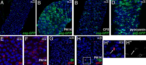
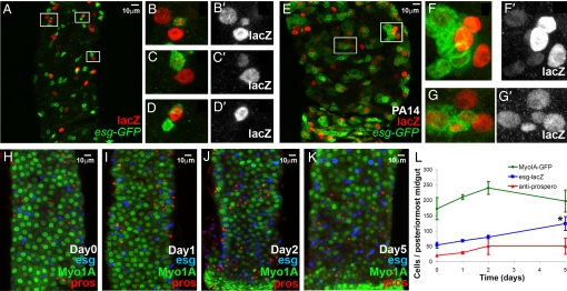
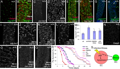
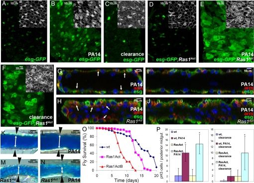
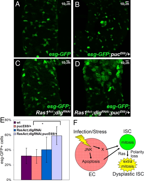
Accumulating evidence suggests that hyperproliferating intestinal stem cells (SCs) and progenitors drive cancer initiation, maintenance, and metastasis. In addition, chronic inflammation and infection have been increasingly recognized for their roles in cancer. Nevertheless, the mechanisms by which bacterial infections can initiate SC-mediated tumorigenesis remain elusive. Using a Drosophila model of gut pathogenesis, we show that intestinal infection with Pseudomonas aeruginosa, a human opportunistic bacterial pathogen, activates the c-Jun N-terminal kinase (JNK) pathway, a hallmark of the host stress response. This, in turn, causes apoptosis of enterocytes, the largest class of differentiated intestinal cells, and promotes a dramatic proliferation of SCs and progenitors that serves as a homeostatic compensatory mechanism to replenish the apoptotic enterocytes. However, we find that this homeostatic mechanism can lead to massive over-proliferation of intestinal cells when infection occurs in animals with a latent oncogenic form of the Ras1 oncogene. The affected intestines develop excess layers of cells with altered apicobasal polarity reminiscent of dysplasia, suggesting that infection can directly synergize with the genetic background in predisposed individuals to initiate SC-mediated tumorigenesis. Our results provide a framework for the study of intestinal bacterial infections and their effects on undifferentiated and mature enteric epithelial cells in the initial stages of intestinal cancer. Assessment of progenitor cell responses to pathogenic intestinal bacteria could provide a measure of predisposition for apoptotic enterocyte-assisted intestinal dysplasias in humans.


Conflict of interest statement

The authors declare no conflict of interest.

Figures

Fig. 1.
Fig. 1.
P. aeruginosa infection induces intestinal progenitor expansion. (A–D) Posterior midgut cells of uninfected (A), strain PA14 infected (B), strain CF5 infected (C) and pyocyanin-fed (D) flies. SCs and progenitors of esg-GAL4 UAS-srcGFP flies fed for 5 days are shown in green. (E and F) Posterior midgut SCs marked with α-Delta antibody (red) of uninfected (E) and PA14-fed (F) flies. (G, H) Posterior midgut SCs marked with α-Delta (green) and phospho-histone-H3 (pH3; red) antibody, of uninfected (G) and PA14-fed (H) flies. (H′ and H″) Magnification of rectangular region in (H), showing colocalization of pH3 (H′; arrow) and Delta (H′ and H″; arrow) staining. Nuclei in all panels are marked with DAPI (blue).
Fig. 2.
Fig. 2.
Mature midgut cells are produced and cellular homeostasis is sustained during infection. (A–K) Lineage tracing of midgut clones induced by esg expressing cells in the absence (A–D) or presence (E–G) of PA14 infection. Green represents GFP driven by esg-GAL4 and red represents nuclear lacZ, the lineage-tracing marker. esg+ cells produce mature enterocytes in both cases (B–D, F, and G), although in the case of PA14 infection, the clone size increases (E–G). (H–L) Time-course of PA14 infection. Myo1A-Gal4 UAS-GFP flies were fed on PA14-containing food for 0 (H), 1 (I), 2 (J), and 5 days (K) and the different cell types [Myo1A+, mature enterocytes (green), esg-lacZ+, SCs and progenitors (blue) and prospero+, enteroendocrine cells (red)] comprising their posterior-most midguts were counted. (L) Only esg+ cell numbers changed significantly during the course of infection (≈2.5-fold increase at day 5, *, P = 0.05; n = 3 intestines).
Fig. 3.
Fig. 3.
P. aeruginosa infection triggers proliferation of SCs and progenitors via JNK-mediated apoptosis of enterocytes. (A and B) Uninfected (A) and PA14-fed (B) midgut enterocytes of esg-GAL4 UAS-srcGFP flies show substantial activation of caspase-3 (red) with concomitant accumulation of GFP (esg+ cells) in their neighboring SCs and progenitors. (C, D) JNK pathway activation in uninfected (C) and PA14-fed (D) midgut cells of puc-lacZ flies. (E and F) Overexpression of either the apoptotic gene reaper (E) or the activated form of the JNK activating kinase HepAct (F) in the all posterior midgut activates caspase-3 (red) in enterocytes neighboring esg+ cells. (G and H) Expression of reaper (G) or HepAct (H) in the progenitor population does not increase their numbers (arrows in H). (I–R) JNK pathway inhibition via overexpression of JNKDN or inhibition of cell death by p35 overexpression in all posterior midgut cells exhibited a reduced number of esg+ cells (P < 0.05, n = 5 intestines, >1,000 cells; I–M), absence of strong apoptotic caspase-3 staining (N–Q) and increased mortality (solid lines in R, P < 0.001, n = 40) following infection compared to the isogenic control flies. Longevity of these flies (dashed lines in R) is not restricted. (S) Model illustrating the JNK-apoptosis cassette and a putative factor X acting in parallel, or sequentially, to induce SC mitosis upon enterocyte (EC) infection or stress.
Fig. 4.
Fig. 4.
Ras1Act oncogene synergizes with infection to induce SC-mediated intestinal dysplasia. (A–F) Control tub-GAL80ts esg-GAL4 UAS-GFP flies (A–C) alone or in combination with UAS-Ras1Act (D–F) that are either uninfected (A and D), PA14-fed (B and E), or cleared from infection (C and F). Midgut esg+ cells are shown in green (A–F) and Arm staining in white in insets. (G–J) Cross-sections (G and H) and sagittal sections (I and J) of PA14-infected wild-type (wt) (G and I) and Ras1Act (H and J) midguts indicating the difference in epithelial structure. DAPI marks the nuclei and red Arm staining. Arm is localized in lateral cell junctions, but in Ras1Act there is mislocalization of Arm in the apical domain (arrows in G and H). (K–N) Posterior midguts of wt (K and L) and Ras1Act (M and N) flies fed on sucrose (K and M) or PA14 (L and N) for 5 days, followed by ingestion of blue dye cornmeal to highlight the gut lumen. White double arrows and black arrowheads delineate the borders of the intestinal epithelium. (O) Control flies survive longer compared to flies expressing two different insertions of Ras1Act (P < 0.001; n = 40). (P) pH3+ cells per posterior midgut are significantly and similarly increased during infection in both control and Ras1Act flies, but differ following clearance (*, P < 0.05; n = 7).
Fig. 5.
Fig. 5.
Synergism between JNK activation and Ras1Act expression and dlg downregulation. Posterior midgut SCs and progenitors marked with GFP (green) from flies of the genotype tub-GAL80ts esg-GAL4 UAS-GFP without (A) or with JNK activation due to the pucE69 mutation (B) or with Ras1Act and dlgRNAi expression (C) or with Ras1Act and dlgRNAi expression in the pucE69 background (D). (E) Ras1Act and dlgRNAi only increased the percent of esg+ cells in pucE69 flies (P < 0.05, n = 5 intestines). (F) Model of the diversion of homeostasis (as described in Fig. S3) toward dysplasia due to Ras and polarity gene alterations causing intestinal SC predisposition.

Similar articles

Cited by

References

    1. Loktionov A. Cell exfoliation in the human colon: Myth, reality, and implications for colorectal cancer screening. Int J Cancer. 2007;120:2281–2289. - PubMed
    1. El-Omar EM. The importance of interleukin 1beta in Helicobacter pylori associated disease. Gut. 2001;48:743–747. - PMC - PubMed
    1. Quante M, Wang TC. Inflammation and stem cells in gastrointestinal carcinogenesis. Physiology (Bethesda) 2008;23:350–359. - PMC - PubMed
    1. Selgrad M, et al. The role of viral and bacterial pathogens in gastrointestinal cancer. J Cell Physiol. 2008;216:378–388. - PMC - PubMed
    1. Lakatos PL, Fischer S, Lakatos L, Gal I, Papp J. Current concept on the pathogenesis of inflammatory bowel disease-crosstalk between genetic and microbial factors: Pathogenic bacteria and altered bacterial sensing or changes in mucosal integrity take “toll”? World J Gastroenterol. 2006;12:1829–1841. - PMC - PubMed

Publication types

MeSH terms

Substances

LinkOut - more resources