. 2009 Dec 15;122(Pt 24):4505-15.
doi: 10.1242/jcs.049155. Epub 2009 Nov 24.

Toll-dependent antimicrobial responses in Drosophila larval fat body require Spätzle secreted by haemocytes

Affiliations

Toll-dependent antimicrobial responses in Drosophila larval fat body require Spätzle secreted by haemocytes

Alice K H Shia et al. J Cell Sci. .

Abstract

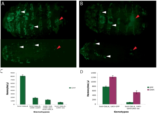
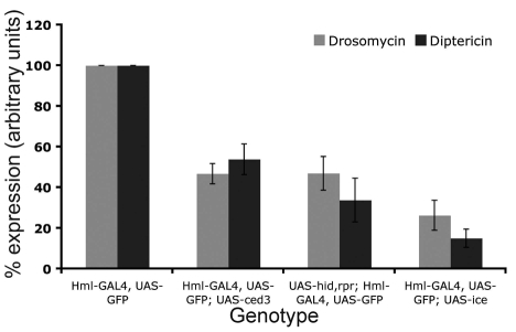
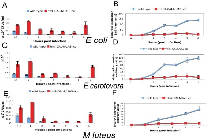
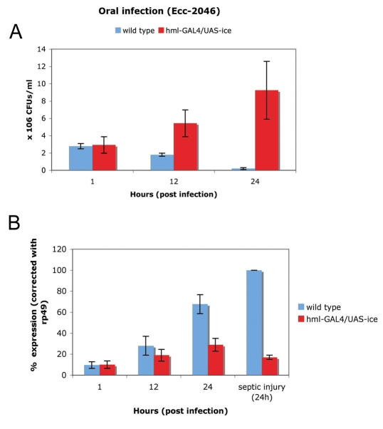
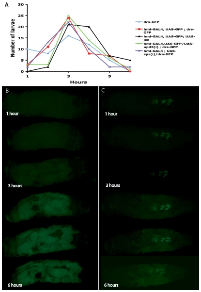
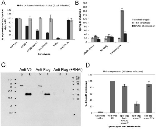
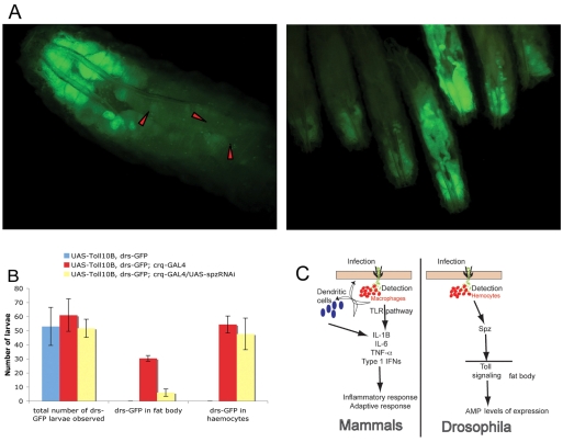
In Drosophila, the humoral response characterised by the synthesis of antimicrobial peptides (AMPs) in the fat body (the equivalent of the mammalian liver) and the cellular response mediated by haemocytes (blood cells) engaged in phagocytosis represent two major reactions that counter pathogens. Although considerable analysis has permitted the elucidation of mechanisms pertaining to the two responses individually, the mechanism of their coordination has been unclear. To characterise the signals with which infection might be communicated between blood cells and fat body, we ablated circulating haemocytes and defined the parameters of AMP gene activation in larvae. We found that targeted ablation of blood cells influenced the levels of AMP gene expression in the fat body following both septic injury and oral infection. Expression of the AMP gene drosomycin (a Toll target) was blocked when expression of the Toll ligand Spätzle was knocked down in haemocytes. These results show that in larvae, integration of the two responses in a systemic reaction depend on the production of a cytokine (spz), a process that strongly parallels the mammalian immune response.


Figures

Fig. 1.
Fig. 1.
Ablation of haemocytes using the GAL4-UAS system. (A) hmlGAl4 and (B) pxnGAL4 were crossed to a UAS line expressing the human apoptotic gene ICE. The cross was hml-GAL4, UAS-GFP or pxn-GAL4, UAS-GFP × TM6B/UAS-ICE (hml-GAL4, UAS-GFP; UAS-ICE /+ or pxn-GAL4, UAS-GFP; UAS-ICE /+, bottom panels of A and B, respectively). Larvae in top panels are the hmlGAL4, UAS-GFP; TM6B/+ and pxnGAL4, UAS-GFP; TM6B/+ siblings of the ablated larvae, hence their Tubby appearance. Visualisation of ablation through a UAS-GFP transgene showed that circulating blood cells and a large number of sessile haemocytes were eliminated (compare top and bottom panels in both A and B). However, some groups of sessile haemocytes were resistant (white arrowheads). The larval haematopoietic organ (red arrowheads) was not deleted but was severely reduced. (C) Quantification of circulating haemocytes marked with GPF in hmlGAL4, UAS-GFP larvae after expression of C elegans ced-3, Drosophila hid and reaper (hid, rpr) and human ICE measured by the number of cells/μl of blood. The hmlGAL4, UAS-GFP strain (without any of the pro-apoptotic gene UAS constructs) was used as the wild-type control (hml). Six independent experiments were performed. The graph represents the mean values ± s.d. Similar results were obtained with pxnGAL4 and srpD-GAL4 (not shown). (D) Comparison between the total number of circulating haemocytes (DAPI) and those labelled by GFP in hmlGAL4, UAS-GFP larvae (left column) or hmlGAL4, UAS-GFP; UAS-ICE larvae (right column). Approximately 59% of circulating haemocytes were deleted by ablation.
Fig. 2.
Fig. 2.
Reduced levels of AMP production by the fat body in haemocyte-ablated larvae. Ablated larvae were infected with Gram-positive bacteria (M. luteus; drosomycin expression used as a read-out for the response and assayed 12 hours post-infection) or Gram-negative bacteria (E. coli; diptericin expression used as a read-out for the response and assayed 6 hours post-infection). AMP expression levels were assayed by northern blot and quantified. Four independent experiments were performed. Mean values ± s.d. are presented. The Pearson correlation test was used to establish whether reduction in AMP expression levels correlated well with the extent of haemocyte ablation (compare Fig. 2 with Fig. 1C). The test found a strong correlation between level of ablation and reduction of drs (P=0.03, r=0.87) or dipt (P=0.02, r=0.88). This reduction was not due to the reduced number of haemocytes producing less AMP because, after quantification in dissected tissue, we found that the fat body accounted for 70% of all AMP gene expression (see Table 1).
Fig. 3.
Fig. 3.
Bacterial persistence versus AMP gene induction in haemocyte-ablated larvae following septic injury. (A,C,E) Proliferation at various times post-infection. In wild-type larvae, bacteria were eliminated within the first hour of infection for E. coli (A) and Ecc (C) or within 2 hours for M. luteus (E). By contrast, haemocyte-depleted larvae failed to efficiently eliminate the bacteria. Although CFUs were reduced within 5 hours post-infection, bacterial proliferation resumed and reached the initial load at the end of the time course (compare blue and red columns in A, C and E at 0.5 and 6 hours post-infection). (B,D,F) Activation of dipt or drs at various times post-infection. Proliferation correlated with a poor activation of dipt (B,D) or drs (F) following infection in haemocyte-ablated larvae (monitored by quantitative real-time PCR). Values in all graphs are mean values ± s.d. of at least three independent experiments. Experiments were done in whole larvae.
Fig. 4.
Fig. 4.
Bacterial persistence versus AMP gene induction in haemocyte-ablated larvae following oral infection. (A) During oral infection, wild-type larvae eliminated Ecc-246 within 24 hours. By contrast, haemocyte-depleted larvae could not restrict bacterial proliferation and exhibited elevated CFUs during the same time course. Despite the higher number of CFUs, systemic induction of dipt (measured by northern blot) was severely reduced in comparison to wild-type larvae (B). This showed that the presence of haemocytes was important for the systemic induction of immunity during oral infection. Mean values ± s.d. from at least three independent experiments are presented (northern blots in whole larvae).
Fig. 5.
Fig. 5.
Fat body expression of drs-GFP in live haemocyte-ablated larvae. (A) Graph showing the distribution of the number of larvae (various genetic backgrounds indicated in colour) initiating expression of drs-GFP in a particular time following immune challenge. As shown, there was essentially no difference between ablated and wild-type larvae in the timing of expression initiation. DD1 is a different chromosomal insertion of drs-GFP used to exclude background specific results. (B,C) Stills from time-lapse imaging of infected wild-type (hmlGAL4/drsGFP) larvae (B) or haemocyte-ablated (hmlGAL4/drsGFP; UAS-ICE larvae) (C) during a 6-hour period. In wild-type larvae, drs-GFP expression initiates and rapidly spreads through the fat body tissue. By contrast, larvae lacking haemocytes do not exhibit the same pattern because initiation of expression occurred but subsequent propagation of expression was not observed. In all frames, posterior is to the left. Similar results were obtained with DD1; hmlGAL4 and DD1; hmlGAL4; UAS-ICE.
Fig. 6.
Fig. 6.
Induction of spz is required in larval haemocytes. (A) Targeting a hairpin construct against spz in haemocytes resulted in the reduction of drs expression levels following infection with M. luteus. A further reduction was observed when hairpin expression was combined with a heterozygous recessive mutant background for spz [spz(i)/rm7] at 25°C or two copies of the hairpin and driver construct [spz(i)/spz(i)] reared at 29°C throughout postembryonic development. Expression of UAS-spz(i) in the fat body [fb; spz(i)] through the lsp2-GAL4 line did not influence drs expression levels. Wild-type levels of drs induction were measured by infecting the GAL4 driver line on its own (hml). Because a short-lived AMP activation occurs through wounding, this was measured by injection of phosphate-buffered saline (buffer). Mean values ± s.d. from three independent experiments are presented. Expression of drs was measured 12 hours following infection. Note that induction of dipt (measured 6 hours following E. coli infection) was not affected by the spz knockdown. (B) To support the above findings we measured activation of spz transcription following infection. This was done by using quantitative real-time PCR following infection with M. luteus. We found that spz was predominantly induced in haemocytes and that this infection-dependent activation was severely reduced using a UAS-RNAi construct expressed in haemocytes (hml-GAL4). (C) A tagged version of spz (spztag) was used to explore the potential function of spz as a signal relayed from blood cells to fat body. Western blots of larval haemolymph showed a clear identification of the Spztag band expressed through hml-GAL4. The quantities loaded for each lane were 5 μg for the Anti-V5 lanes and 10 μg for anti-FLAG and anti-FLAG (+RNAi) lanes, respectively. Concomitant expression of a UAS-spzRNAi transgene with the UAS-spztag (anti-FLAG +RNAi lane) sufficiently silenced the latter at the protein level. N, non-reduced samples; R, reduced samples. Concentrations of the antibodies used were 1:10000 for Anti-V5 and 1:500 for Anti-FLAG. (D) Expression of the UAS-spztag using the hml-GAL4 in spz-deficient background (spzrm7) rescued drs induction following infection by septic injury (drs induction assayed by northern blots in whole larvae). Mean values ± s.d. from at least three independent experiments are presented.
Fig. 7.
Fig. 7.
Expression of Toll10B in larval haemocytes induces drs-GFP in fat body. (A) Expression of UAS-Toll10B using the haemocyte-specific crq-GAL4 (Silva et al., 2007) in axenic conditions of culture induced drs-GFP in both the haemocytes (red arrowheads in magnified larva, left panel) as well as the fat body (right panel; note that the expression was variable but characteristically involved activation at the posterior). (B) Statistical representation of the data, where the number of UAS-Toll10B, drs-GFP; crq-GAL4 larvae expressing GFP in the fat body was significantly reduced when UAS-spzRNAi was included in the configuration. Mean values ± s.d. are from three independent crosses of UAS-Toll10B, drs-GFP with crq-GAL4 in axenic conditions. The total number of third instar larvae observed for each experiment was n1=40, n2=52, n3=67 for UAS-Toll10B, drs-GFP; n1=48, n2=65, n3=70 for UAS-Toll10B, drs-GFP; crq-GAL4 and n1=47, n2=49, n3=59 for UAS-Toll10B, drs-GFP; crq-GAL4/UAS-RNAi. (C) Schematic model for cytokine release in mammals (macrophages) and Drosophila (haemocytes), following an infection. TRL, Toll-like receptor; IL, interleukin; TNF, tumor necrosis factor; IFN, interferon.

Similar articles

Cited by

References

    1. Agaisse, H., Petersen, U. M., Boutros, M., Mathey-Prevot, B. and Perrimon, N. (2003). Signalling role of haemocytes in Drosophila JAK/STAT-dependent response to septic injury. Dev. Cell 5, 441-450. - PubMed
    1. Akira, S., Uematsu, S. and Takeuchi, O. (2006). Pathogen recognition and innate immunity. Cell 124, 783-801. - PubMed
    1. Anderson, K. V. and Nüsslein-Volhard, C. (1984). Genetic analysis of dorsal-ventral embryonic pattern in Drosophila. In Pattern Formation: A Primer in Developmental Biology (ed. G. Malacinski and S. V. Bryant), pp. 269-289. New York: Macmillan.
    1. Anderson, K. V., Jürgens, G., Nüsslein-Volhard, C. (1985) Establishment of dorsal-ventral polarity in the Drosophila embryo: genetic studies on the role of the Toll gene product. Cell 42, 779-789. - PubMed
    1. Basset, A., Khush, R. S., Braun, A., Gardan, L., Boccard, F., Hoffmann, J. A. and Lemaitre, B. (2000). The phytopathogenic bacteria Erwinia carotovora infects Drosophila and activates an immune response. Proc. Natl. Acad. Sci. USA 97, 3376-3381. - PMC - PubMed

Publication types

MeSH terms