. 2010 Jan;38(3):832-45.
doi: 10.1093/nar/gkp1039. Epub 2009 Nov 24.

SIRT1 deacetylates APE1 and regulates cellular base excision repair

Affiliations

SIRT1 deacetylates APE1 and regulates cellular base excision repair

Tohru Yamamori et al. Nucleic Acids Res. 2010 Jan.

Abstract

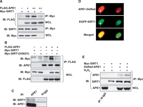
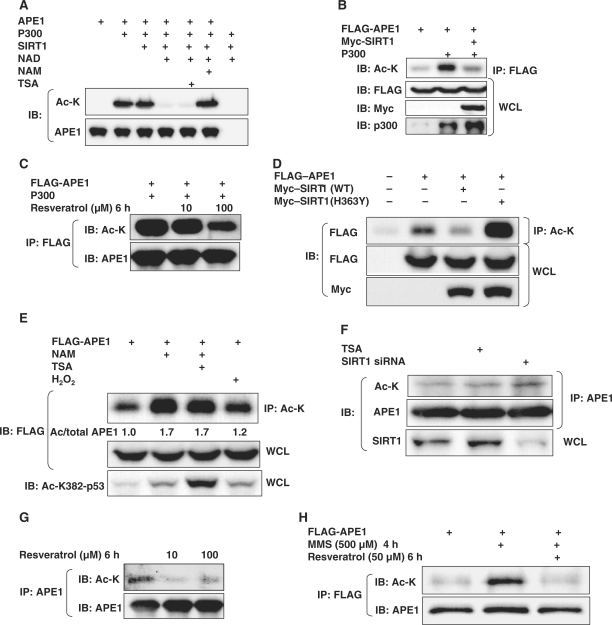
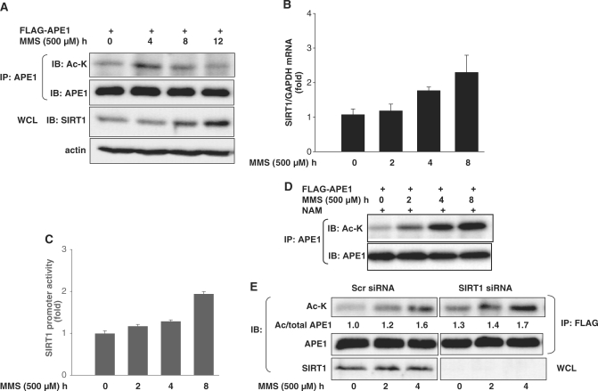
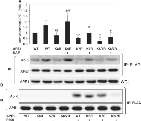
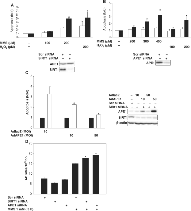
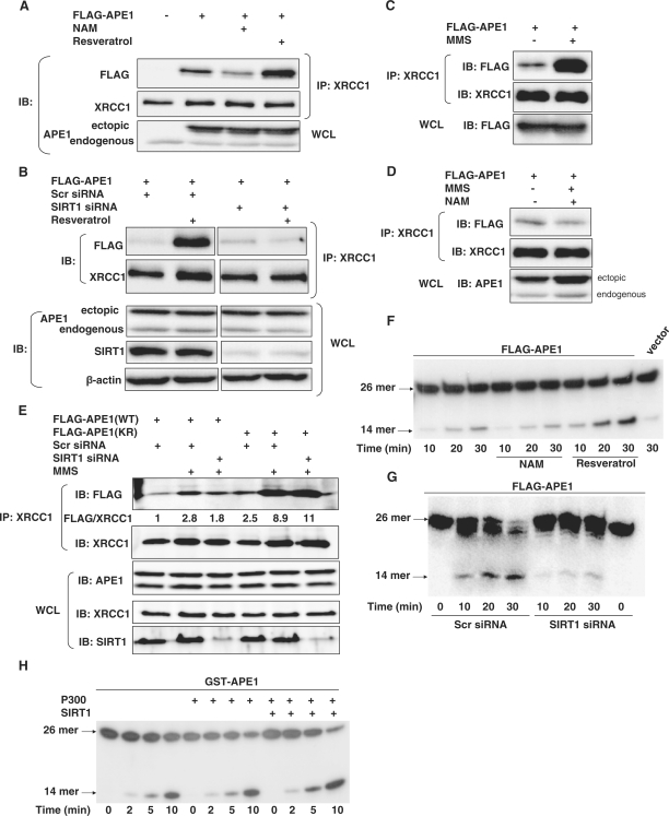
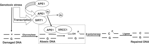
Apurinic/apyrimidinic endonuclease-1 (APE1) is an essential enzyme in the base excision repair (BER) pathway. Here, we show that APE1 is a target of the SIRTUIN1 (SIRT1) protein deacetylase. SIRT1 associates with APE1, and this association is increased with genotoxic stress. SIRT1 deacetylates APE1 in vitro and in vivo targeting lysines 6 and 7. Genotoxic insults stimulate lysine acetylation of APE1 which is antagonized by transcriptional upregulation of SIRT1. Knockdown of SIRT1 increases cellular abasic DNA content, sensitizing cells to death induced by genotoxic stress, and this vulnerability is rescued by overexpression of APE1. Activation of SIRT1 with resveratrol promotes binding of APE1 to the BER protein X-ray cross-complementing-1 (XRCC1), while inhibition of SIRT1 with nicotinamide (NAM) decreases this interaction. Genotoxic insult also increases binding of APE1 to XRCC1, and this increase is suppressed by NAM or knockdown of SIRT1. Finally, resveratrol increases APE activity in XRCC1-associated protein complexes, while NAM or knockdown of SIRT1 suppresses this DNA repair activity. These findings identify APE1 as a novel protein target of SIRT1, and suggest that SIRT1 plays a vital role in maintaining genomic integrity through regulation of the BER pathway.


Figures

Figure 1.
Figure 1.
APE1 and SIRT1 associate with each other. (A) APE1 and SIRT1 bind to each other in HEK 293 cells. Immunoprecipitation of epitope-tagged SIRT1 co-precipitates epitope-tagged APE1 expressed in HEK 293 cells. (B) APE1 does not bind to catalytically inactive dominant negative SIRT1. Immunoprecipitation of epitope-tagged APE1 co-precipitates epitope-tagged wild-type SIRT1 but not dominant negative SIRT1 (H363Y) expressed in HEK 293 cells. (C) Endogenous SIRT1 binds to endogenous APE1. Co-immunoprecipitation of endogenous SIRT1 and APE1 in HEK 293 cells. (D) Co-localization of fluorescent epitope-tagged SIRT1 and APE1 expressed in HEK 293 cells. Co-localization of extra-nucleolar (white arrow) APE1 but not nucleolar (blue arrow) APE1 with SIRT1 is shown. (E) Hydrogen peroxide (H2O2) promotes binding of APE1 to SIRT1. H2O2 (500 µM, 30 min) increases co-precipitation of epitope-tagged SIRT1 in immunoprecipitates of epitope-tagged APE1 expressed in HEK 293 cells. WCL: whole cell lysate. N-IgG: non-immune immunoglobulin. FLAG-APE1 and Myc-SIRT1 were expressed in A and B, DsRed-APE1 and EGFP-SIRT1 in C, and DsRed-APE1 and Myc-SIRT1 in D.
Figure 2.
Figure 2.
SIRT1 deacetylates APE1. (A) In vitro deacetylation of recombinant APE1 by SIRT1. Recombinant APE1 enzymatically acetylated by the p300 acetyltransferase was deacetylated by recombinant active SIRT1 in presence of the SIRT1 co-factor NAD+ (1 mM). Deacetylation was inhibited by the SIRT1 inhibitor nicotinamide (NAM, 10 mM), but not the class I and II HDAC inhibitor Trichostatin A (TSA, 1 µM). (B) APE1 acetylated by the p300 acetyltransferase is deacetylated by SIRT1 in vivo. (C) Resveratrol inhibits p300-induced lysine acetylation of exogenous APE1. (D) Expression of wild-type SIRT1 (WT) decreases and dominant negative SIRT1 (H363Y) increases, acetylation of exogenous APE1 in HEK293 cells. (E) The SIRT1 inhibitor NAM and H2O2 increases acetylation of exogenous APE1 in HEK 293 cells. Cells were treated with H2O2 (500 µM), NAM (5 mM) and the class I and II HDAC inhibitor Trichostatin A (TSA: 5 µM) for 30 min. Values of lysine acetylated/total APE1 from a representative experiment, normalized to untreated cells, are shown. Acetylation of endogenous p53 is shown at bottom for comparison. WCL: whole cell lysate. (F) TSA (5 µM) and siRNA-mediated knockdown of endogenous SIRT1 increase lysine acetylation of endogenous APE1 in HEK 293 cells. (G) Resveratrol suppresses basal lysine acetylation of APE1 in HEK 293 cells. (H) Activation of endogenous SIRT1 with resveratrol antagonizes MMS-induced lysine acetylation of exogenous APE1.
Figure 3.
Figure 3.
SIRT1 is upregulated by MMS and antagonizes MMS-induced acetylation of APE1. (A) Reciprocal temporal relationship between MMS-induced APE1 acetylation and SIRT1 expression. Time course of APE1 lysine acetylation and SIRT1 protein expression in HEK 293 cells challenged with MMS. (B) MMS increases SIRT1 mRNA. Time course of SIRT1 mRNA upregulation in HeLa cells challenged with MMS. (C) MMS increases SIRT1 promoter activity. Time course of human SIRT1 promoter induction in HeLa cells challenged with MMS. (D) SIRT1 inhibition augments MMS-induced acetylation of APE1. Time course of APE1 lysine acetylation in HEK 293 cells pre-treated with the SIRT1 inhibitor NAM (5 mM) and challenged with MMS. (E) SiRNA-mediated suppression of SIRT1 in HEK 293 cells promotes MMS-induced APE1 acetylation. Lysine acetylated/total APE1 ratio from a representative experiment is shown and is normalized to untreated control siRNA cells. WCL: whole cell lysate.
Figure 4.
Figure 4.
SIRT1 targets lysines 6 and 7 in APE1 for deacetylation. (A) Basal lysine acetylation, and increase in lysine acetylation by treatment with the SIRT1 inhibitor nicotinamide (NAM: 5 mM, 16 h), of epitope-tagged wild-type and non-acetylatable mutants of APE1 expressed in HEK 293 cells. Lysine acetylated/total APE1 was quantified and is expressed relative to wild-type APE1 in untreated cells. *P < 0.05 and #P > 0.05 compared with untreated cells transfected with the same construct. ##P > 0.05, and **P < 0.005 compared with untreated cells transfected with WT APE1. ###P > 0.05, ***P < 0.05, and ΨP < 0.005 compared with NAM-treated cells transfected with WT APE1. N = 4 in all conditions. (B) Targeting of the N-terminal lysines in APE1 for acetylation by the p300 acetyltransferase. Epitope-tagged wild-type (WT) APE1, or the non-acetylatable lysine mutants of APE1, were expressed in HEK 293 cells, with and without overexpression of the p300 acetyltransferase. WCL: whole cell lysate.
Figure 5.
Figure 5.
SIRT1 protects from genotoxic stress-induced cell death through APE1. Apoptotic death with oxidative stress (H2O2 for 24 h) and abasic DNA damage (MMS for 24 h) in HeLa cells in which SIRT1 expression (A) or APE1 expression (B) is knocked down with SIRT1 or APE1 siRNA (black bars). Scrambled (scr) siRNA (white bars) was used as control. Apoptosis is expressed as fold change compared with untreated cells. *P < 0.05 and **P < 0.01 compared with control siRNA. Knockdown of SIRT1 and APE1 is shown at bottom. (C) APE1 overexpression rescues cells with SIRT1 down-regulation from MMS-induced apoptosis. Apoptotic cell death induced by MMS (200 µM, 24 h) in HeLa cells treated with SIRT1 siRNA (white bars) or scrambled (scr) siRNA (black bars) were infected with a control virus (AdLacZ) that expresses the inert E. coli LacZ gene, or and adenovirus that expresses APE1 (AdAPE1). Apoptosis is expressed as fold change compared with control siRNA. *P < 0.05 compared with cells infected with AdLacZ. Knockdown of SIRT1 and adenoviral overexpression of APE1 is shown at right. (D) SIRT1 plays a role in abasic DNA repair. SIRT1 or APE1 was knocked down in HeLa cells with siRNA. Apurinic/apyrimidinic DNA sites were quantified in untreated cells and cells treated with MMS for 3 h. *P < 0.05 compared with control siRNA.
Figure 6.
Figure 6.
SIRT1 promotes cellular endonuclease activity toward abasic DNA sites by stimulating the binding of APE1 to XRCC1. (A) Endogenous SIRT1 promotes binding of APE1 to XRCC1. Co-immunoprecipitation of endogenous XRCC1 with epitope-tagged APE1 expressed in HeLa cells treated with the SIRT1 inhibitor NAM (5 mM), or the SIRT1 activator resveratrol (50 µM) for 6 h. (B) Resveratrol-stimulated association between APE1 and XRCC1 is mediated by SIRT1. Co-immunoprecipitation of epitope-tagged APE1 and endogenous XRCC1 in HeLa cells, with and without resveratrol (50 µM, 6 h), that are treated with scrambled siRNA (Scr siRNA) or SIRT1 siRNA. Separated panels are from the same immunoblot, with irrelevant lanes deleted. (C) Genotoxic stress promotes binding of APE1 to XRCC1. Co-immunoprecipitation of epitope-tagged APE1 and endogenous XRCC1 in HeLa cells treated with MMS (500 µM, 6 h) (D) SIRT1 mediates genotoxic stress-induced binding of APE1 to XRCC1. Co-immunoprecipitation of epitope-tagged APE1 and endogenous XRCC1 in HeLa cells treated with MMS (500 µM, 6 h) with and without treatment with SIRT1 inhibitor NAM (5 mM). (E) SIRT1 mediates genotoxic stress-stimulated binding of wild-type, but not acetylation-deficient APE1. Co-immunoprecipitation of endogenous XRCC1 and epitope-tagged wild-type APE1 (WT) or mutated APE1 (KR) that is non-acetylatable on lysines 6 and 7, in HeLa cells treated with MMS (500 µM, 6 h), with or without knockdown of SIRT1. Cells were co-transfected with scr siRNA or SIRT1 siRNA. WCL: whole cell lysate. (F and G) SIRT1 promotes AP endonuclease activity in XRCC1-bound protein complex. AP endonuclease activity toward abasic DNA in XRCC1 immunoprecipitates from (F) HeLa cells expressing FLAG-tagged APE1 and treated with the SIRT1 activator resveratrol (50 µM), or the SIRT1 inhibitor NAM (5 mM) for 6 h and (G) HeLa cells in which SIRT1 expression is knocked down with siRNA. (H) P300-induced acetylation, and SIRT1-induced deacetylation, of recombinant APE1 does not affect its AP endonuclease activity. In vitro AP endonuclease activity toward abasic DNA of recombinant GST-APE1 that is acetylated by the p300 acetyltransferase, followed by deacetylation by SIRT1.
Figure 7.
Figure 7.
Scheme depicting the role of SIRT1 in the BER of damaged DNA. Rectangle shows the AP endonuclease step. Ac: acetyl.

Similar articles

Cited by

References

    1. Demple B, Harrison L. Repair of oxidative damage to DNA: enzymology and biology. Annu. Rev. Biochem. 1994;63:915–948. - PubMed
    1. Izumi T, Wiederhold LR, Roy G, Roy R, Jaiswal A, Bhakat KK, Mitra S, Hazra TK. Mammalian DNA base excision repair proteins: their interactions and role in repair of oxidative DNA damage. Toxicology. 2003;193:43–65. - PubMed
    1. Mitra S, Izumi T, Boldogh I, Bhakat KK, Hill JW, Hazra TK. Choreography of oxidative damage repair in mammalian genomes. Free Radic. Biol. Med. 2002;33:15–28. - PubMed
    1. Tell G, Damante G, Caldwell D, Kelley MR. The intracellular localization of APE1/Ref-1: more than a passive phenomenon? Antioxid. Redox Signal. 2005;7:367–384. - PubMed
    1. Bennett RA, Wilson DM, 3rd, Wong D, Demple B. Interaction of human apurinic endonuclease and DNA polymerase beta in the base excision repair pathway. Proc. Natl Acad. Sci. USA. 1997;94:7166–7169. - PMC - PubMed

Publication types

MeSH terms