. 2009 Dec 1;15(23):7246-55.
doi: 10.1158/1078-0432.CCR-09-1292. Epub 2009 Nov 24.

Mesenchymal stem cell carriers protect oncolytic measles viruses from antibody neutralization in an orthotopic ovarian cancer therapy model

Affiliations

Mesenchymal stem cell carriers protect oncolytic measles viruses from antibody neutralization in an orthotopic ovarian cancer therapy model

Emily K Mader et al. Clin Cancer Res. .

Abstract

Purpose: Preexisting antiviral antibodies in cancer patients can quickly neutralize oncolytic measles virus (MV) and decrease its antitumor potency. In contrast to "naked" viruses, cell-associated viruses are protected from antibody neutralization. Hence, we hypothesized that measles virotherapy of ovarian cancer in measles-immune mice might be superior if MV-infected mesenchymal stem cell (MSC) carriers are used.

Experimental design: Antimeasles antibodies titers in ovarian cancer patients were determined. The protection of MV by MSC from antimeasles antibodies, the in vivo biodistribution profiles, and tumor infiltration capability of MSC were determined. Measles-naïve or immune tumor-bearing mice were treated with naked virus or MSC-associated virus and mice survivals were compared.

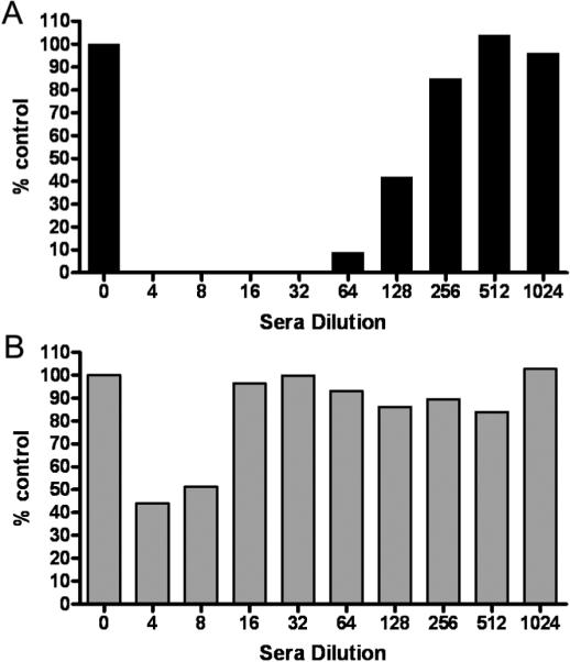
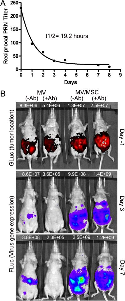
Results: MSC transferred MV infection to target cells via cell-to-cell heterofusion and induced syncytia formation in the presence of high titers of antimeasles antibody, at levels that completely inactivated naked virus. Athymic mice bearing i.p. human SKOV3ip.1 ovarian tumor xenografts passively immunized with measles-immune human serum were treated with saline, naked MV, or MV-infected MSC. Bioluminescent and fluorescent imaging data indicated that i.p. administered MSC localized to peritoneal tumors, infiltrated into the tumor parenchyma, and transferred virus infection to tumors in measles naïve and passively immunized mice. Survival of the measles-immune mice was significantly enhanced by treatment with MV-infected MSC. In contrast, survivals of passively immunized mice were not prolonged by treatment with naked virus or uninfected MSC.

Conclusions: MSC should be used as carriers of MV for intraperitoneal virotherapy in measles-immune ovarian cancer patients.


Figures

Figure 1
Figure 1
Adipose tissue derived mesenchymal stem cells (MSC) are susceptible to infection by measles virus. (A) Photographs MV-RFP infected MSC taken at 48h post infection using different multiplicities of infection (MOI). Cells were maintained in the absence (−FIP) or presence (+FIP) of a fusion inhibitory peptide, FIP. (B) Quantitation of MV-RFP infection in MSC by analyzing for numbers of RFP positive cells (+FIP) by flow cytometry at 48h post infection. (C) Viability of infected MSC over time. (D) Progeny produced by virus infected MSC or Vero cells at 48h post infection at MOI 0.2 or 1.0. Amount of cell-associated virus or released virus in the supernatant were titrated on Vero cells. Error bars represent S.D. (n=3 replicates).
Figure 2
Figure 2
Virus delivered by infected MSC carriers is more resistant to inhibition by antimeasles antibodies than by cell-free `naked' virus. (A) MV-GFP virus was mixed with varying dilutions of human measles immune sera (1:4 to 1:1024 dilution in 5% FBS-DMEM) and added to Vero cells. (B) MSC were infected with MV-GFP (MOI 1.0) and the next day, MSC were mixed with varying dilutions of human measles immune sera and overlaid on Vero cells. The Vero cells were cultured for 2 days and the numbers of syncytia were counted and represented as a percentage of numbers of syncytia found in wells with no serum. A representative experiment from three replicates is shown.
Figure 3
Figure 3
In vivo distribution of MSC post IP delivery. (A) Serial bioluminescent imaging of mice post IP injection of MV-Luc infected MSC (MV/MSC) into mice with no tumor or mice bearing intraperitoneal human SKOV3ip.1 ovarian tumors. Another group of tumor-bearing mice received MV-Luc (MV). (B) MV infected MSC were labeled with a fluorescent CellTracker Red CMTPX dye and injected IP into mice bearing peritoneal SKOV3ip.1, A2780 or OVCAR5 human ovarian tumors. Mice were euthanized 24 hours later. Photographs (40X or 100X magnification) of freshly harvested tissues showing red fluorescent MSCs on the omentum or peritoneal wall of mice with no tumor or on the tumor nodules of mice. (C) Confocal microscopy images of tumor cryosections demonstrating presence of red fluorescent MSC on the surface and in the parenchyma of SKOV3ip.1 tumors. Scale bars represent 100 μm unless otherwise indicated.
Figure 4
Figure 4
MSC protect measles virus from complete neutralization by anti-measles antibody in mice. (A) Timeline for decay of human measles immune serum in sera of athymic mice over time. Mice were euthanized at each time point to obtain serum for virus plaque reduction neutralization (PRN) assays. Each point is the average of PRN assays from 2 to 4 mice. Curve fit R2=0.99. (B) Representative photographs showing correlation between tumor location (indicated by GLuc activity) and MV-Luc gene expression (FLuc activity), delivered by cell-free virus or MSC-associated virus in measles naïve or measles immune mice. Mice were imaged using colenterazine substrate for GLuc expression one day before imaging for FLuc activity using D-luciferin substrate. The photons count (photons/sec) for each mouse is as indicated.
Figure 5
Figure 5
MSC mediated delivery of measles virus enhanced survival of measles immune mice. (A) Kaplan Meier survival curves of mice given different treatments; saline, uninfected MSC, 105 TCID50 `naked' MV-NIS or 105 MV-NIS infected MSC (MOI 4.0, ~ 60% infection) in measles naïve mice (−Ab) or measles immune mice (+Ab, 100 EU/mouse). (B) Immunohistochemical staining for measles-nucleocapsid (MV-N) protein (blue staining=site of viral gene expression) and CD68 positive macrophages (brown/red staining). Arrows in the last panel point to corresponding necrotic area in the tumor with minimal MV-N staining but strong CD68 staining. Scale bars indicate 500 μm.

References

    1. Jemal A, Siegel R, Ward E, et al. Cancer statistics, 2008. CA Cancer J Clin. 2008;58:71–96. - PubMed
    1. Badgwell D, Bast RC., Jr. Early detection of ovarian cancer. Dis Markers. 2007;23:397–410. - PMC - PubMed
    1. Fung-Kee-Fung M, Oliver T, Elit L, et al. Optimal chemotherapy treatment for women with recurrent ovarian cancer. Curr Oncol. 2007;14:195–208. - PMC - PubMed
    1. Rocconi RP, Numnum TM, Stoff-Khalili M, et al. Targeted gene therapy for ovarian cancer. Curr Gene Ther. 2005;5:643–653. - PubMed
    1. Kimball KJ, Numnum TM, Rocconi RP, Alvarez RD. Gene therapy for ovarian cancer. Curr Oncol Rep. 2006;8:441–447. - PubMed

Publication types