

Invasion of human breast cancer cells in vivo requires both paracrine and autocrine loops involving the colony-stimulating factor-1 receptor
- PMID: 19934330
- PMCID: PMC2794986
- DOI: 10.1158/0008-5472.CAN-09-1868
Invasion of human breast cancer cells in vivo requires both paracrine and autocrine loops involving the colony-stimulating factor-1 receptor
Abstract
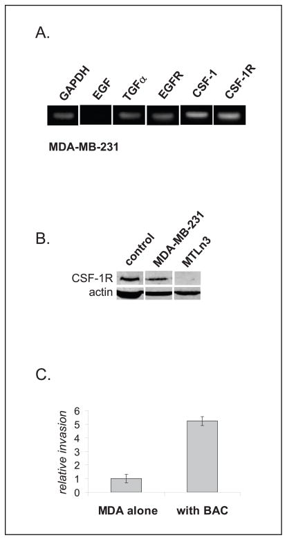
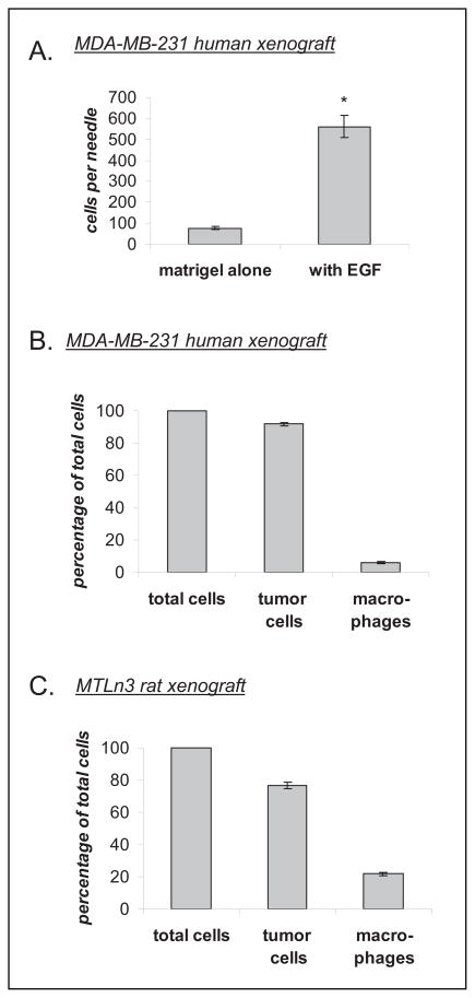
Colony-stimulating factor-1 (CSF-1) and its receptor (CSF-1R) have been implicated in the pathogenesis and progression of various types of cancer, including breast cancer. This is based on high levels of circulating CSF-1 in patient sera with aggressive disease and increased CSF-1R staining in the tumor tissues. However, there have been no direct in vivo studies to determine whether a CSF-1 autocrine signaling loop functions in human breast cancer cells in vivo and whether it contributes to invasion. Recently, in mouse and rat models, it has been shown that invasion and metastasis are driven by an epidermal growth factor (EGF)/CSF-1 paracrine loop between tumor cells and host macrophages. In this macrophage-dependent invasion, tumor cells secrete CSF-1 and sense EGF, whereas the macrophages secrete EGF and sense CSF-1. Here, we test the hypothesis that in human breast tumors, the expression of both the CSF-1 ligand and its receptor in tumor cells leads to a CSF-1/CSF-1R autocrine loop which contributes to the aggressive phenotype of human breast tumors. Using MDA-MB-231 cell-derived mammary tumors in severe combined immunodeficiency mice, we show here for the first time in vivo that invasion in a human mammary tumor model is dependent on both paracrine signaling with host macrophages as well as autocrine signaling involving the tumor cells themselves. In particular, we show that the autocrine contribution to invasion is specifically amplified in vivo through a tumor microenvironment-induced upregulation of CSF-1R expression via the transforming growth factor-beta1.
Figures


Similar articles
-
Macrophages promote the invasion of breast carcinoma cells via a colony-stimulating factor-1/epidermal growth factor paracrine loop.Cancer Res. 2005 Jun 15;65(12):5278-83. doi: 10.1158/0008-5472.CAN-04-1853. Cancer Res. 2005. PMID: 15958574
-
Autocrine CSF1R signaling mediates switching between invasion and proliferation downstream of TGFβ in claudin-low breast tumor cells.Oncogene. 2015 May 21;34(21):2721-31. doi: 10.1038/onc.2014.226. Epub 2014 Aug 4. Oncogene. 2015. PMID: 25088194 Free PMC article.
-
The EGF/CSF-1 paracrine invasion loop can be triggered by heregulin beta1 and CXCL12.Cancer Res. 2009 Apr 1;69(7):3221-7. doi: 10.1158/0008-5472.CAN-08-2871. Epub 2009 Mar 17. Cancer Res. 2009. PMID: 19293185 Free PMC article.
-
CSF-1 and its receptor in breast carcinomas and neoplasms of the female reproductive tract.Mol Reprod Dev. 1997 Jan;46(1):71-4. doi: 10.1002/(SICI)1098-2795(199701)46:1<71::AID-MRD11>3.0.CO;2-6. Mol Reprod Dev. 1997. PMID: 8981366 Review.
-
The role of CSF-1 in normal physiology of mammary gland and breast cancer: an update.Exp Biol Med (Maywood). 2004 Jan;229(1):1-11. doi: 10.1177/153537020422900101. Exp Biol Med (Maywood). 2004. PMID: 14709771 Review.
Cited by
-
Direct visualization of the phenotype of hypoxic tumor cells at single cell resolution in vivo using a new hypoxia probe.Intravital. 2016;5(2):e1187803. doi: 10.1080/21659087.2016.1187803. Epub 2016 May 16. Intravital. 2016. PMID: 27790387 Free PMC article.
-
Migratory gene expression signature predicts poor patient outcome: are cancer stem cells to blame?Breast Cancer Res. 2012 Nov 15;14(6):114. doi: 10.1186/bcr3338. Breast Cancer Res. 2012. PMID: 23153392 Free PMC article.
-
In Vivo Visualization of Stromal Macrophages via label-free FLIM-based metabolite imaging.Sci Rep. 2016 May 25;6:25086. doi: 10.1038/srep25086. Sci Rep. 2016. PMID: 27220760 Free PMC article.
-
Blood vessel endothelium-directed tumor cell streaming in breast tumors requires the HGF/C-Met signaling pathway.Oncogene. 2017 May 11;36(19):2680-2692. doi: 10.1038/onc.2016.421. Epub 2016 Nov 28. Oncogene. 2017. PMID: 27893712 Free PMC article.
-
Autocrine CSF-1 and CSF-1 receptor coexpression promotes renal cell carcinoma growth.Cancer Res. 2012 Jan 1;72(1):187-200. doi: 10.1158/0008-5472.CAN-11-1232. Epub 2011 Nov 3. Cancer Res. 2012. PMID: 22052465 Free PMC article.
References
-
- Condeelis J, Pollard JW. Macrophages: obligate partners for tumor cell migration, invasion, and metastasis. Cell. 2006;124:263–6. - PubMed
-
- Wyckoff JB, Wang Y, Lin EY, et al. Direct visualization of macrophage-assisted tumor cell intravasation in mammary tumors. Cancer Res. 2007;67:2649–56. - PubMed
-
- Wyckoff J, Wang W, Lin EY, et al. A paracrine loop between tumor cells and macrophages is required for tumor cell migration in mammary tumors. Cancer Res. 2004;64:7022–9. - PubMed
-
- Goswami S, Sahai E, Wyckoff JB, et al. Macrophages promote the invasion of breast carcinoma cells via a colony-stimulating factor-1/epidermal growth factor paracrine loop. Cancer Res. 2005;65:5278–83. - PubMed
Publication types
MeSH terms
Substances
Grants and funding
LinkOut - more resources
Full Text Sources
Medical
Molecular Biology Databases
Research Materials
Miscellaneous
