

In vitro osteogenic differentiation of adipose stem cells after lentiviral transduction with green fluorescent protein
- PMID: 19934675
- PMCID: PMC2862472
- DOI: 10.1097/SCS.0b013e3181bf04af
In vitro osteogenic differentiation of adipose stem cells after lentiviral transduction with green fluorescent protein
Abstract
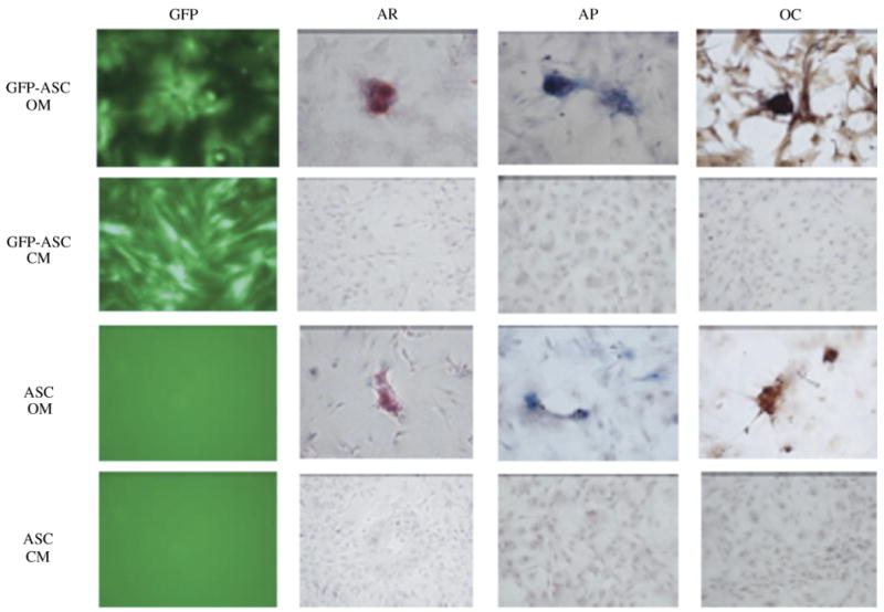
Background: Adipose-derived stem cells (ASCs) have the potential to differentiate into osteogenic cells that can be seeded into scaffolds for tissue engineering for use in craniofacial bone defects. Green fluorescent protein (GFP) has been widely used as a lineage marker for mammalian cells. The use of fluorescent proteins enables cells to be tracked during manipulation such as osteogenic differentiation within three-dimensional scaffolds. The purpose of this study was to examine whether ASCs introduced with GFP-encoding lentivirus vector exhibit adequate GFP fluorescence and whether the expression of GFP interfered with osteogenic differentiation of ASCs in both monolayer and three-dimensional scaffolds in vitro.
Methods: Primary ASCs were harvested from the inguinal fat pad of Sprague Dawley rats. Isolated ASCs were cultured and infected with a lentiviral vector encoding GFP and plated into both monolayers and three-dimensional scaffolds in vitro. The cells were then placed in osteogenic medium. Osteogenic differentiation of the GFP-ASCs was assessed using alizarin red S, alkaline phosphate staining, and immunohistochemistry staining of osteocalcin with quantification of alizarin red S and osteocalcin staining.
Results: The efficacy of infection of ASCs with a lentiviral vector encoding GFP was high. Cell-cultured GFP-ASCs remained fluorescent over the 8 weeks of the study period. The GFP-ASCs were successfully induced into osteogenic cells both in monolayers and three-dimensional scaffolds. Whereas the quanitification of alizarin red S revealed no difference between osteoinduced ASCs with or without GFP, the quantification of osteocalcin revealed increased staining in the GFP group.
Conclusions: Transduction of isolated ASCs using a lentiviral vector encoding GFP is an effective method for tracing osteoinduced ASCs in vitro. Quantification data showed no decrease in staining of the osteoinduced ASCs.
Figures


References
-
- Langer R, Vacanti JP. Tissue engineering. Science. 1993;260:920. - PubMed
-
- Dudas JR, Marra KG, Cooper GM, et al. The osteogenic potential of adipose-derived stem cells for the repair of rabbit calvarial defects. Ann Plast Surg. 2006;56:543. - PubMed
-
- Kokai LE, Rubin JP, Marra KG. The potential of adipose-derived adult stem cells as a source of neuronal progenitor cells. Plast Reconstr Surg. 2005;116:1453. - PubMed
-
- Lee JH, Kemp DM. Human adipose-derived stem cells display myogenic potential and perturbed function in hypoxic conditions. Biochem Biophys Res Commun. 2006;341:882. - PubMed
MeSH terms
Substances
Grants and funding
LinkOut - more resources
Full Text Sources
