

Opioid-induced decreases in rat brain adenosine levels are reversed by inhibiting adenosine deaminase
- PMID: 19934879
- PMCID: PMC2784661
- DOI: 10.1097/ALN.0b013e3181bdf894
Opioid-induced decreases in rat brain adenosine levels are reversed by inhibiting adenosine deaminase
Abstract
Background: Opioids disrupt sleep and adenosine promotes sleep, but no studies have characterized the effects of opioids on adenosine levels in brain regions known to regulate states of arousal. Delivering opioids to the pontine reticular formation (PRF) and substantia innominata (SI) region of the basal forebrain disrupts sleep. In contrast, administering adenosine agonists to the PRF or SI increases sleep. These findings encouraged the current study testing the hypothesis that microdialysis delivery of opioids to the PRF or SI decreases adenosine levels in the PRF or SI, respectively.
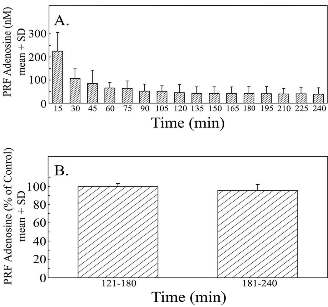
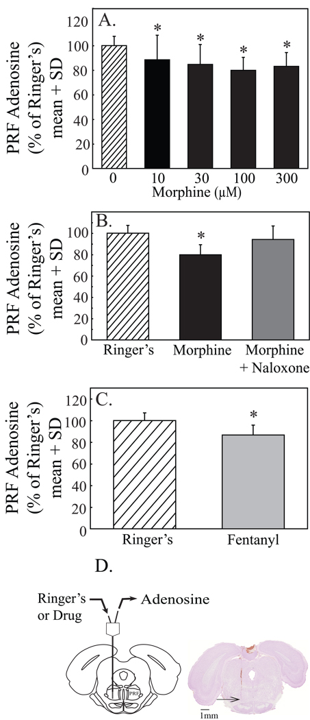
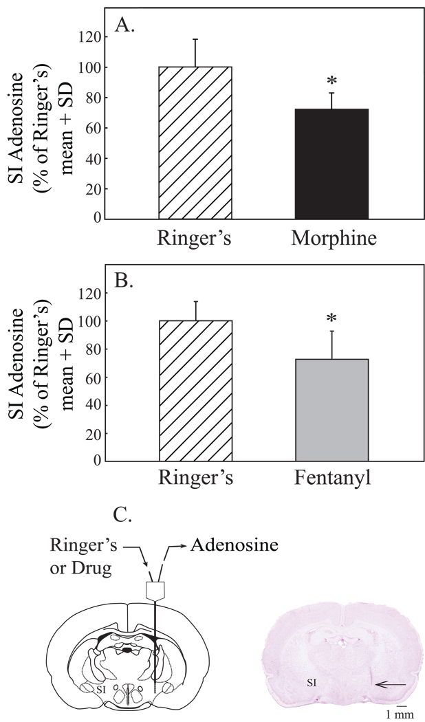
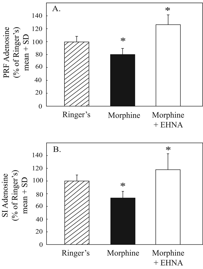
Methods: A microdialysis probe was placed in the PRF of isoflurane anesthetized rats and perfused with Ringer's solution (control) followed by Ringer's solution containing morphine (0, 10, 30, 100, or 300 microm), fentanyl (100 microm), morphine (100 microm) and the adenosine deaminase inhibitor EHNA (100 microm), or naloxone (10 microm) and morphine (100 microm). Additional experiments measured adenosine levels in the SI before and during microdialysis delivery of morphine, fentanyl, and morphine plus EHNA.
Results: Morphine caused a significant (P < 0.05) concentration-dependent decrease in PRF adenosine levels. The significant decrease (-20%) in adenosine caused by 100 microm morphine was blocked by coadministration of naloxone. Fentanyl also significantly decreased (-13.3%) PRF adenosine. SI adenosine levels were decreased by morphine (-26.8%) and fentanyl (-27.4%). In both PRF and SI, coadministration of morphine and EHNA prevented the significant decrease in adenosine levels caused by morphine alone.
Conclusions: These data support the interpretation that decreased adenosine levels in sleep-regulating brain regions may be one of the mechanisms by which opioids disrupt sleep.
Conflict of interest statement
Figures
Comment in
-
Opiates, sleep, and pain: the adenosinergic link.Anesthesiology. 2009 Dec;111(6):1175-6. doi: 10.1097/ALN.0b013e3181bdfa2e. Anesthesiology. 2009. PMID: 19934853 Free PMC article. No abstract available.
References
-
- Lydic R, Baghdoyan HA. In: Neurochemical mechanisms mediating opioid-induced REM sleep disruption, Sleep and Pain. Lavigne G, Sessle BJ, Choinière M, Soja PJ, editors. Seattle: International Association for the Study of Pain (IASP) Press; 2007. pp. 99–122.
-
- Roehrs T, Roth T. Sleep and pain: Interaction of two vital functions. Semin Neurol. 2005;25:106–116. - PubMed
-
- Kundermann B, Krieg JC, Schreiber W, Lautenbacher S. The effect of sleep deprivation on pain. Pain Res Manag. 2004;9:25–32. - PubMed
-
- Gallagher RM, Rosenthal LJ. Chronic pain and opiates: Balancing pain control and risks in long-term opioid treatment. Arch Phys Med Rehabil. 2008;89:S77–S82. - PubMed
Publication types
MeSH terms
Substances
Grants and funding
LinkOut - more resources
Full Text Sources
Other Literature Sources
Medical
Molecular Biology Databases
Research Materials
