Case Reports
. 2009 Dec;65(6 Suppl):226-36.
doi: 10.1227/01.NEU.0000350868.95634.CA.

Preoperative sensorimotor mapping in brain tumor patients using spontaneous fluctuations in neuronal activity imaged with functional magnetic resonance imaging: initial experience

Affiliations
Case Reports

Preoperative sensorimotor mapping in brain tumor patients using spontaneous fluctuations in neuronal activity imaged with functional magnetic resonance imaging: initial experience

Dongyang Zhang et al. Neurosurgery. 2009 Dec.

Abstract

Objective: To describe initial experience with resting-state correlation mapping as a potential aid for presurgical planning of brain tumor resection.

Methods: Resting-state blood oxygenation-dependent functional magnetic resonance imaging (fMRI) scans were acquired in 17 healthy young adults and 4 patients with brain tumors invading sensorimotor cortex. Conventional fMRI motor mapping (finger-tapping protocol) was also performed in the patients. Intraoperatively, motor hand area was mapped using cortical stimulation.

Results: Robust and consistent delineation of sensorimotor cortex was obtained using the resting-state blood oxygenation-dependent data. Resting-state functional mapping localized sensorimotor areas consistent with cortical stimulation mapping and in all patients performed as well as or better than task-based fMRI.

Conclusion: Resting-state correlation mapping is a promising tool for reliable functional localization of eloquent cortex. This method compares well with "gold standard" cortical stimulation mapping and offers several advantages compared with conventional motor mapping fMRI.


Figures

Figure 1
Figure 1
Resting state correlation maps showing the distribution of the sensorimotor network in normal subjects. A: The average correlation map in a group of 17 healthy young adults. The bilateral pattern represents correlated neuronal activity between left and right sensorimotor cortices. Seed placement is shown by the blue circle. B: The consistency of resting state correlation mapping is demonstrated by repeated scans in single subjects. Four 7 minute scans were performed in each of the 17 subjects. Two subjects are displayed here showing the correlation map from each of the four scans (transverse slices, z=51). Correlations contralateral to the seed are consistently seen in the same region within the sensorimotor network in each of the repeated scans. All images are displayed left-on-left (neurologic convention).
Figure 2
Figure 2
Case 1. A: Structural MRI showing the location of the tumor which primarily disrupts right parietal, somatosensory and motor cortices (neurologic convention). The green circle represents the location of ipsilateral hand response to cortical stimulation. B: Resting state correlation map showing the distribution of the sensorimotor network in this patient. A seed was placed in the sensorimotor cortex of the contralateral hemisphere (blue circle). Correlations in the ipsilateral hemisphere were predominantly localized to a region anterior and lateral to the tumor, displaced by the intracranial mass. All images are displayed left-on-left (neurologic convention).
Figure 3
Figure 3
Comparison of resting state and task-related fMRI mapping in Case 1. A: Finger-tapping fMRI × 2. Activity within the tumor (blue arrows) was seen in trial 2 but not in trial 1. B: Resting state correlation mapping × 2 shows a similar distribution of correlated activity, resembling the activation from trial 1 but not trial 2 of the task. All images are displayed left-on-left (neurologic convention).
Figure 4
Figure 4
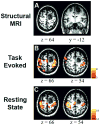
Case 2. A: Structural MRI revealed a tumor in right parietal cortex extending anteriorly into motor and somatosensory areas (neurologic convention). B: Task-related fMRI failed to reveal activity within the sensorimotor cortex. A deliberately low threshold reveals noise outside of the brain without responses within the sensorimotor network. C: Resting state correlation mapping using a seed in contralateral hemisphere (same seed as in Figure 2B, blue circle) showed that ipsilateral sensorimotor cortex near the tumor was displaced anteriorly and laterally. All images are displayed left-on-left (neurologic convention).
Figure 5
Figure 5
Case 3. A: Structural MRI revealed a tumor in left parietal cortex that invades territory near the central sulcus (neurologic convention). The green circle represents the location of ipsilateral hand response to cortical stimulation. B: Task-related activity was seen bilaterally in frontal lobe. In addition, a large band of activity appeared in right parietal cortex, not consistent with the pattern of activity from the sensorimotor network. C: Resting state correlation mapping using a seed in the right (unaffected) hemisphere (blue circle) showed ipsilateral correlations anterior to the tumor as well as a region of activity in midline parietal cortex. Note absence in the correlation mapping results of parietal activity seen in the task-related map. D: ICA decomposition is an alternative method that does not rely on a priori seed placement. Results from ICA verify the anterior shift of the sensorimotor network. E. Parietal activation seen during task-evoked scan is revealed to be a separate resting state network that is normally dissociated from the sensorimotor network (seed: blue circle). All images are displayed left-on-left (neurologic convention).
Figure 6
Figure 6
Case 4. A: Structural MRI showed a mass in right frontal cortex (neurologic convention). The green circle represents the location of ipsilateral hand response to cortical stimulation. B: Task-related mapping shows activity within the sensorimotor network but also small responses in parietal cortex that are seemingly unrelated to motor function or sensation. C: Resting state correlation mapping showed that the sensorimotor network was largely unaffected by the tumor anterior to the central sulcus. The seed region is marked by the blue circle. All images are displayed left-on-left (neurologic convention).
Figure 7
Figure 7
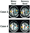
Separation of Sensory and Motor Activity. Motor and sensory cortices exhibit highly correlated neuronal activity. Separation of activity unique to each area can be achieved using partial correlation. Two seeds are defined, representing motor and sensory cortex (blue circles). Partial correlation eliminates activity that is shared between the two ROIs before correlation maps are computed. Shown for two representative cases, correlations contralateral to the seeds (blue box) can be seen to segregate along the central sulcus. The location and angle of the central sulcus is approximated by the blue diagonal. All images are displayed left-on-left (neurologic convention).

References

    1. Adcock JE, Wise RG, Oxbury JM, Oxbury SM, Matthews PM. Quantitative fMRI assessment of the differences in lateralization of language-related brain activation in patients with temporal lobe epilepsy. Neuroimage. 2003;18:423–438. - PubMed
    1. Arfanakis K, Cordes D, Haughton VM, Moritz CH, Quigley MA, Meyerand ME. Combining independent component analysis and correlation analysis to probe interregional connectivity in fMRI task activation datasets. Magn Reson Imaging. 2000;18:921–930. - PubMed
    1. Beckmann CF, DeLuca M, Devlin JT, Smith SM. Investigations into resting-state connectivity using independent component analysis. Philosophical transactions of the Royal Society of London. 2005;360:1001–1013. - PMC - PubMed
    1. Behrens TE, Woolrich MW, Jenkinson M, Johansen-Berg H, Nunes RG, Clare S, Matthews PM, Brady JM, Smith SM. Characterization and propagation of uncertainty in diffusion-weighted MR imaging. Magn Reson Med. 2003;50:1077–1088. - PubMed
    1. Binder JR, Swanson SJ, Hammeke TA, Morris GL, Mueller WM, Fischer M, Benbadis S, Frost JA, Rao SM, Haughton VM. Determination of language dominance using functional MRI: a comparison with the Wada test. Neurology. 1996;46:978–984. - PubMed

Publication types

MeSH terms