Review
. 2009 Dec;9(12):874-85.
doi: 10.1038/nrc2761.

Mucins in cancer: function, prognosis and therapy

Affiliations
Review

Mucins in cancer: function, prognosis and therapy

Donald W Kufe. Nat Rev Cancer. 2009 Dec.

Abstract

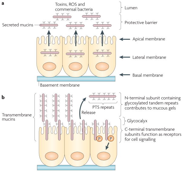
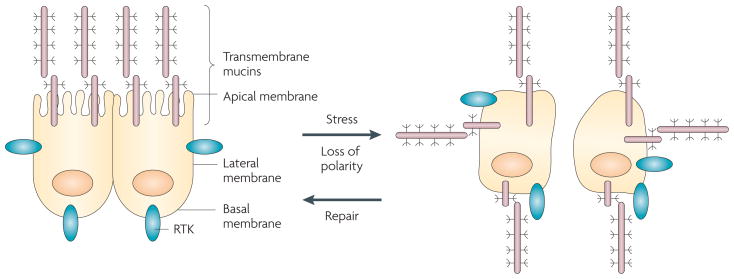
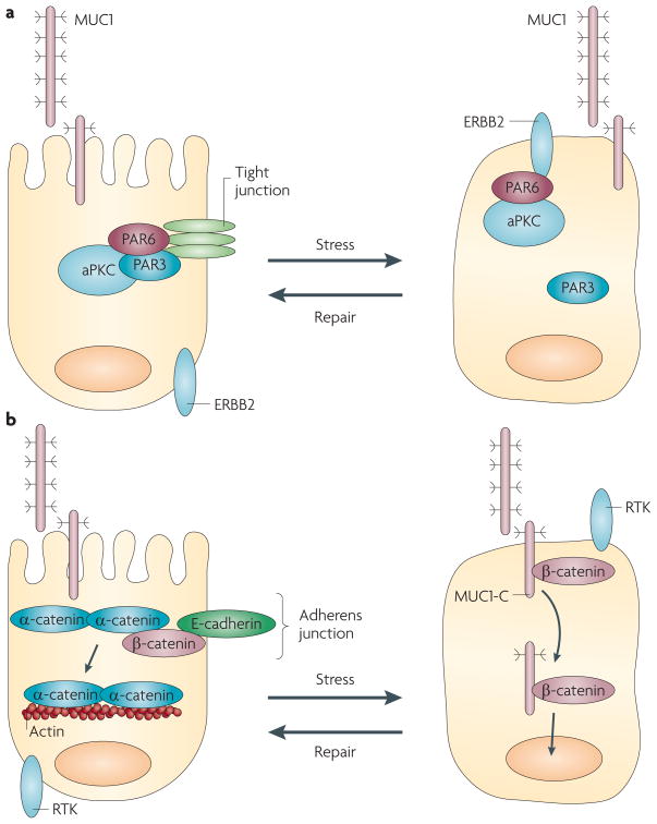
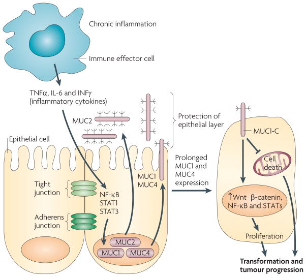
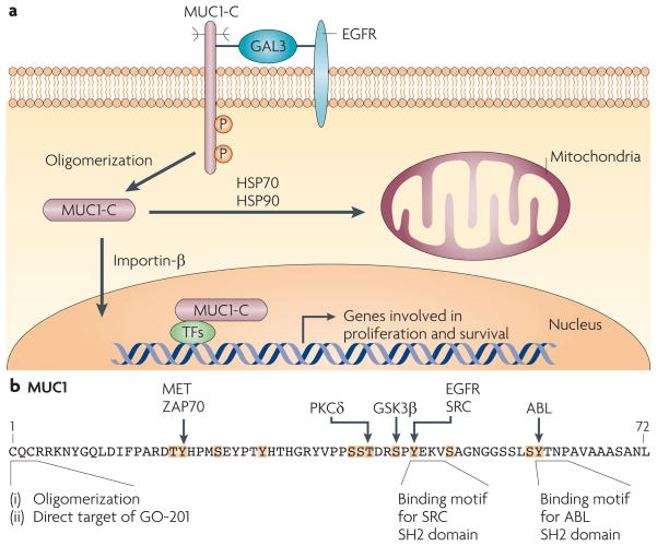
Epithelia are protected from adverse conditions by a mucous barrier. The secreted and transmembrane mucins that constitute the mucous barrier are largely unrecognized as effectors of carcinogenesis. However, both types of mucins are intimately involved in inflammation and cancer. Moreover, diverse human malignancies overexpress transmembrane mucins to exploit their role in signalling cell growth and survival. Mucins have thus been identified as markers of adverse prognosis and as attractive therapeutic targets. Notably, the findings that certain transmembrane mucins induce transformation and promote tumour progression have provided the experimental basis for demonstrating that inhibitors of their function are effective as anti-tumour agents in preclinical models.


Conflict of interest statement

Competing interests statement

The author declares competing financial interests: see web version for details.

Figures

Figure 1
Figure 1. Secreted and transmembrane mucins form physical barriers that protect epithelia
a| The secreted mucins are released from the apical membrane to form a protective gel that limits exposure to commensal bacteria and suppresses the inflammatory response. The gel also protects the epithelial layer from adverse conditions, for example exposure to ingested toxins, reactive oxygen species (ROS) and proteolytic enzymes in the gastrointestinal tract. Mucin 2 (MUC2) is the major secreted mucin lining the gastrointestinal mucosa. b | The transmembrane mucins are expressed in the apical cell membrane so that the region containing the glycosylated tandem proline, threonine and serine (PTS) repeats extends as a rigid structure beyond the glycocalyx and into the mucous gel. MUC1 and MUC4, and possibly MUC13, are heterodimers that are translated as single polypeptides and cleaved into amino- and carboxy-terminal subunits that in turn form a stable non-covalent complex. The N-terminal mucin subunits containing the PTS repeats are tethered to the cell surface in a complex with the C-terminal transmembrane subunit. Release of the N-terminal subunits into the mucous gel leaves the transmembrane subunits as receptors to signal the presence of inflammation and other forms of stress to the interior of the cell. Evolution of the secreted mucins to include a transmembrane component thus provided an additional level of defence to promote the growth, repair and survival of epithelial cells. MUC16 can also undergo autocleavage; however, it is not known whether MUC16 is expressed as a heterodimeric complex or whether release of the mucin region occurs after positioning of MUC16 in the apical membrane.
Figure 2
Figure 2. Epithelial stress response is associated with loss of polarity and repositioning of cell surface receptors
Epithelial cells are polarized with the separation of the apical and basolateral membranes by specialized tight junctions between neighbouring cells. In contrast to apical positioning of the transmembrane mucins, certain other cell surface molecules, for example the receptor tyrosine kinases (RTKs), are restricted to the basolateral membranes. In the presence of inflammation and other settings that induce damage, there is loss of tight junction integrity and cell polarity. In turn, the transmembrane mucins are repositioned over the entire cell membrane and interact with RTKs. Loss of polarity allows epithelial cells to activate a programme of repair and survival. Whereas the epithelial stress response is reversible and polarity is re-established after repair, loss of polarity is irreversible in carcinoma cells. Therefore, mucin 1 (MUC1) constitutively interacts with diverse RTKs and promotes their downstream signals, thus providing a mechanism by which carcinoma cells can exploit a physiological stress response for their own growth and survival.
Figure 3
Figure 3. Transmembrane mucins, loss of polarity and disruption of cell–cell adhesion
a| Tight junctions are dependent on a complex of the PAR6, PAR3 and atypical protein kinase C (aPKC) proteins. Activation of ERBB2, which is necessary for the epithelial stress response, disrupts the interaction of PAR3 with PAR6 and aPKC, and thereby induces loss of polarity. In carcinoma cells, mucin 1 (MUC1) interacts with ERBB2 and other members of the Erbb family, and promotes ERBB2 signalling. In this setting, constitutive activation of ERBB2 contributes to the irreversible disruption of tight junctions and the loss of polarity that is associated with transformation. b | In adherens junctions, β-catenin links α-catenin to E-cadherin and, in turn, α-catenin forms homodimers and interacts with the actin cytoskeleton. The MUC1 carboxy-terminal transmembrane subunit (MUC1-C) cytoplasmic domain binds to β-catenin and localizes with it in the nucleus, thus sequestering it away from adherens junctions. In addition, overexpression of MUC1 is associated with downregulation of E-cadherin expression, which disrupts adherens junctions. Moreover, β-catenin is sequestered in MUC4–ERBB2 complexes at the apical membrane, which also contributes to the disruption of adherens junction function. RTK, receptor tyrosine kinase.
Figure 4
Figure 4. Mucins, chronic inflammation and cancer
In this proposed model of the association of mucins with chronic inflammation and cancer, the production of inflammatory cytokines by immune effector cells activates transcription factors, for example nuclear factor-κB (NF-κB), signal transducer and activator of transcription 1 (STAT1) and STAT3, in epithelial cells. In turn, these transcription factors upregulate mucin expression to enhance the mucous barrier and protect the epithelial layer. Mucin 2 (MUC2) limits the inflammatory response at the apical membrane and inhibits transformation. Upregulation of the MUC1 and MUC4 transmembrane mucins similarly contributes to the protective barrier and loss of polarity in the epithelial stress response. Activation of MUC1 is associated with targeting of the MUC1 C-terminal transmembrane subunit (MUC1-C) to the nucleus, where it promotes a gene programme for proliferation and survival. Targeting of MUC1-C to the mitochondria also blocks cell death to prevent loss of the epithelial barrier. However, with chronic inflammation and prolonged stimulation of this protective response, epithelial cells may become susceptible to the accumulation of genetic mutations that induce transformation in a setting with downregulation of pathways that would otherwise protect against oncogenic events. IL-6, interleukin-6; IFNγ, interferon-γ; TNFα, tumour necrosis factor-α
Figure 5
Figure 5. Activation of MUC1-C in stress and transformation
a| The mucin 1 (MUC1) C-terminal transmembrane subunit (MUC1-C) forms complexes with the epidermal growth factor receptor (EGFR) that are mediated at least in part by galectin 3 (GAL3) lattices. MUC1-C also forms complexes with ERBB2–4 and with other receptor tyrosine kinases (not shown), although it is not known whether these interactions require galectin 3. MUC1-C–RTK complexes are transiently formed in non-malignant epithelial cells in response to inflammatory cytokines and are constitutively formed in carcinoma cells. MUC1-C oligomers are targeted to the nucleus by an importin-β-dependent mechanism, where MUC1-C associates with several transcription factors (TFs). MUC1-C thereby promotes the transcription of genes that encode proteins involved in proliferative and pro-survival signals by affecting the recruitment of co-activators and co-repressors, and/or regulating the latency of transcription factor occupancy. MUC1-C is also targeted to the mitochondrial outer membrane by heat shock protein 70 (HSP70) and HSP90, where it blocks stress-induced loss of the mitochondrial transmembrane potential,, and the induction of cell death in response to DNA damage, reactive oxygen species, hypoxia and glucose deprivation,–. b | MUC1-C intracellular signalling is mediated in part by phosphorylation of the cytoplasmic domain, which contains five documented and seven potential phosphorylation sites (highlighted in orange with known kinases indicated). The CQC sequence is necessary for oligomerization of MUC1-C and targeting of MUC1-C to the nucleus and mitochondria, as well as binding of MUC1-C to diverse effectors, such as nuclear factor-κB. Direct targeting of the MUC1-C CQC sequence with GO-201 induces complete regression of established human breast and prostate tumour xenografts in mice,. GSK3β, glycogen synthase kinase 3β; P, phosphorylation; PKCδ, protein kinase Cδ; SH2, Src homology 2.

References

    1. Desseyn JL, et al. Evolutionary history of the 11p15 human mucin gene family. J Mol Evol. 1998;46:102–106. - PubMed
    1. Desseyn JL, Aubert JP, Porchet N, Laine A. Evolution of the large secreted gel-forming mucins. Mol Biol Evol. 2000;17:1175–1184. - PubMed
    1. Bork P, Patthy L. The SEA module: a new extracellular domain associated with O-glycosylation. Protein Sci. 1995;4:1421–1425. - PMC - PubMed
    1. Asker N, Axelsson MA, Olofsson SO, Hansson GC. Dimerization of the human MUC2 mucin in the endoplasmic reticulum is followed by a N-glycosylation-dependent transfer of the mono- and dimers to the Golgi apparatus. J Biol Chem. 1998;273:18857–18863. - PubMed
    1. Herrmann A, et al. Studies on the “insoluble” glycoprotein complex from human colon. Identification of reduction-insensitive MUC2 oligomers and C-terminal cleavage. J Biol Chem. 1999;274:15828–15836. - PubMed

Publication types