. 2010 Feb 25;29(8):1103-13.
doi: 10.1038/onc.2009.402. Epub 2009 Nov 23.

Primary ex vivo cultures of human fallopian tube epithelium as a model for serous ovarian carcinogenesis

Affiliations

Primary ex vivo cultures of human fallopian tube epithelium as a model for serous ovarian carcinogenesis

K Levanon et al. Oncogene. .

Abstract

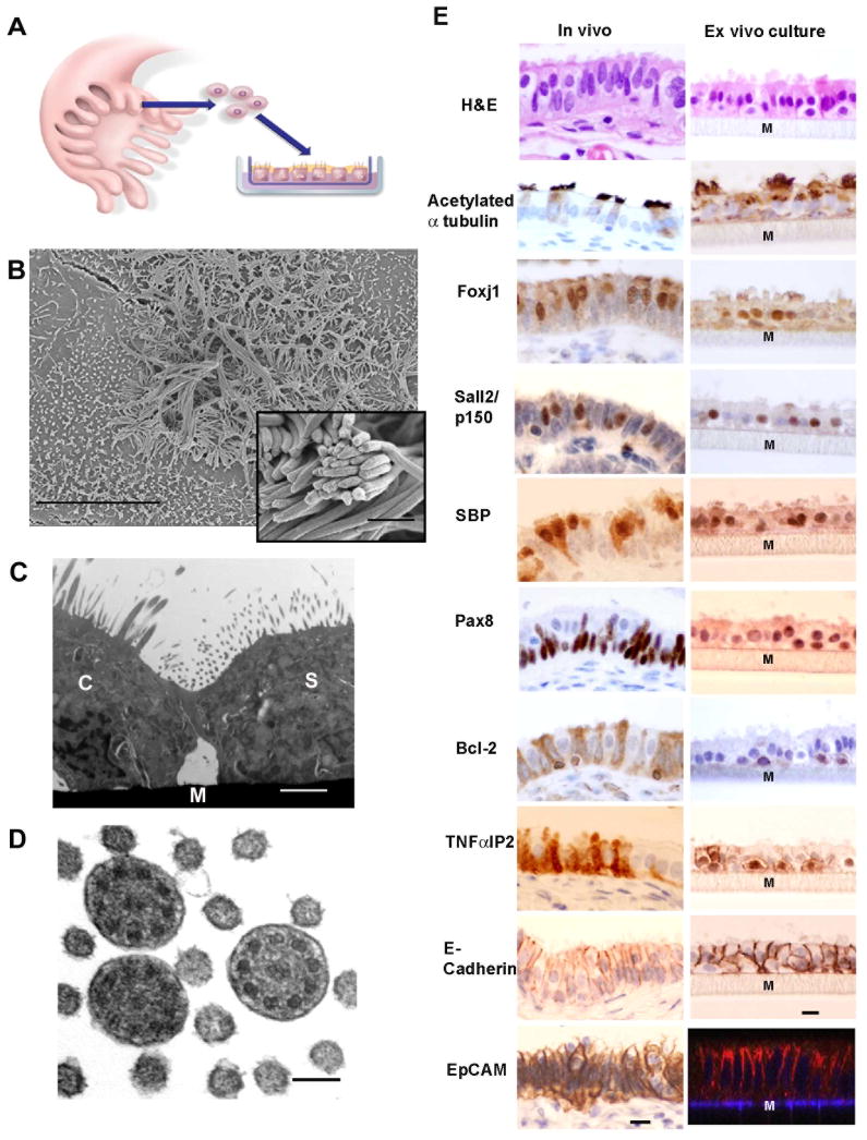
Recent studies suggest that some serous ovarian carcinomas (SOCs) arise from the fallopian tube (FT) epithelium rather than the ovarian surface epithelium. This hypothesis places emphasis on the FT secretory epithelial cell as a cell-of-origin. Herein, we report the development of a novel ex vivo primary human FT epithelium culture system that faithfully recapitulates the in vivo epithelium, as shown by morphological, ultrastructural and immunophenotypic analyses. Mass spectrometry-based proteomics reveal that these cultures secrete proteins previously identified as biomarkers for ovarian cancer. We also use this culture system to study the response of the FT epithelium to genotoxic stress and find that the secretory cells exhibit a distinct response to DNA damage when compared with neighboring ciliated cells. The secretory cells show a limited ability to resolve the damage over time, potentially leaving them more susceptible to accumulation of additional mutagenic injury. This divergent response is confirmed with in situ studies using tissue samples, further supporting the use of this ex vivo culture system to investigate FT epithelial pathobiology. We anticipate that this novel culture system will facilitate the study of SOC pathogenesis, and propose that similar culture systems could be developed for other organ site-specific epithelia.


Figures

Figure 1
Figure 1
The FTE ex-vivo culture system displays morphology and immunomarkers of both FTSECs and ciliated cells. (A) A schematic illustration of the ex-vivo co-culture system. The co-culture is composed of primary human FTE cells grown in the air-liquid interface on Transwell inserts. The cells form a polarized monolayer. (B) Scanning electron microscope image of a confluent culture showing both ciliated cells and secretory cells with microvilli (bar 10μm). Inset shows cilia in higher power (bar 1 μm). (C) Transmission electron microscope image showing a ciliated cell (C) and a secretory cell (S) and the Transwell membrane (M) (bar 1μm). (D) transmission electron microscopy cross section through cilia demonstrating the 9+2 arrangement of axial microtubules (bar 100nm). (E) H&E and immunohistochemistry of normal human fallopian tube fimbria and cross sections of formalin-fixed-paraffin-embedded ex-vivo cultures with the ciliated lineage markers: Acetylated α-tubulin, FoxJ1, p150/Sall2, SELENBP1 (SBP); secretory lineage markers: Pax-8, Bcl-2 and TNFαIP2; pan-epithelial surface markers: E-Cadherin and Ep-CAM (immunofluorescence, Z-stack reconstitution of confocal microscopy images). The gray matrix beneath the cell layer represents the Transwell membrane (Marked by M). Bar 10 μm.
Figure 2
Figure 2. Wound healing in the FT ex-vivo culture system
(A–C) Bright field images of the healing of a scratch in a confluent ex-vivo culture, at day 0 (A), day 1 (B) and day 2 (C) post scratching (red lines mark the borders of the scratch). Proliferating cells on the perimeter of the healing wound, as demonstrated by IF staining for either the proliferation factor Ki67 (D, green) or BrdU (E, green) which co-localize with nuclei of secretory cells (Pax-8 positive nuclei, red) giving a yellow signal. (F) While both cell types exist in the culture, he wound is ‘healed’ by predominantly FT secretory cells (right of the white line), as demonstrated by co-staining for the lineage markers for secretory cells (Pax-8, red) and for ciliated cells (Foxj1, green).
Figure 3
Figure 3. FT ex-vivo culture secretes known FT and ovarian carcinoma proteins
(A) Detection of oviductin (MUC9, ~150KD) and Glycodelin A (PP14, ~28KD) by western blot in conditioned medium of FTE from seven different donors grown in ex-vivo cultures, while there is no detectable protein secretion when grown in regular culture conditions. (B) Detection of HE4 (~28KD), an ovarian cancer serum biomarker, in some established ovarian cancer cell lines and some FTE ex-vivo cultures.
Figure 4
Figure 4. Differential dsDNA break response in the fallopian tube epithelium ex-vivo
(A) Immunofluorescence (en-face images) of representative cells in the ex-vivo culture system, stained for the secretory lineage marker Pax8 (red) and γH2A.X (green), and the nuclei are outlined with DAPI (blue). The formation of γH2A.X foci was recorded 2, 4, 10, 24 and 48 hours after exposure of the co-cultures to ionizing radiation (500 rad). γH2A.X foci are preferentially detectable in FTSECs 2 hours after irradiation and persist for at least 48 hours. Original magnification X60. (B) The kinetics of dsDNA break pathway activation in FTSECs vs. ciliated cells ex-vivo.γH2A.X foci form in FTSECs early after exposure to ionizing radiation, and persist in 70% of the cells for at least 48 hours. Under the same conditions, γH2A.X foci formation in ciliated cells peaks after 10 hours and returns to baseline within 24 hours. The graph represents integration of 4 experiments.
Figure 5
Figure 5. Preferential activation of the dsDNA break checkpoint pathway in FTSEC in-situ
(A) H&E and immunohistochemistry of normal human fallopian tube fimbria following exposure to ionizing radiation, demonstrating preferential activation of the dsDNA break signaling pathway in secretory cells (marked by arrowheads), compared to ciliated cells (marked by arrows). Original magnification ×60. (B) Quantitative analysis of number of adjacent secretory cells and ciliated cells that stained positive for γH2A.X foci and p53 24 hours after exposure to IR in-situ (125–250 rad). No staining was seen in non-irradiated cell.

References

    1. Ando H, Kobayashi M, Toda S, Kikkawa F, Masahashi T, Mizutani S. Establishment of a ciliated epithelial cell line from human Fallopian tube. Hum Reprod. 2000;15:1597–603. - PubMed
    1. Bartkova J, Horejsi Z, Koed K, Kramer A, Tort F, Zieger K, et al. DNA damage response as a candidate anti-cancer barrier in early human tumorigenesis. Nature. 2005;434:864–70. - PubMed
    1. Bast RC, Jr, Badgwell D, Lu Z, Marquez R, Rosen D, Liu J, et al. New tumor markers: CA125 and beyond. Int J Gynecol Cancer. 2005;15(Suppl 3):274–81. - PubMed
    1. Bowen NJ, Logani S, Dickerson EB, Kapa LB, Akhtar M, Benigno BB, et al. Emerging roles for PAX8 in ovarian cancer and endosalpingeal development. Gynecol Oncol. 2007;104:331–7. - PubMed
    1. Branzei D, Foiani M. Regulation of DNA repair throughout the cell cycle. Nat Rev Mol Cell Biol. 2008;9:297–308. - PubMed

Publication types

Substances