. 2009 Nov;5(11):e1000735.
doi: 10.1371/journal.pgen.1000735. Epub 2009 Nov 20.

Caenorhabditis elegans HIM-18/SLX-4 interacts with SLX-1 and XPF-1 and maintains genomic integrity in the germline by processing recombination intermediates

Affiliations

Caenorhabditis elegans HIM-18/SLX-4 interacts with SLX-1 and XPF-1 and maintains genomic integrity in the germline by processing recombination intermediates

Takamune T Saito et al. PLoS Genet. 2009 Nov.

Abstract

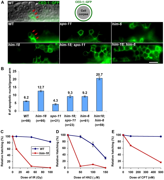
Homologous recombination (HR) is essential for the repair of blocked or collapsed replication forks and for the production of crossovers between homologs that promote accurate meiotic chromosome segregation. Here, we identify HIM-18, an ortholog of MUS312/Slx4, as a critical player required in vivo for processing late HR intermediates in Caenorhabditis elegans. DNA damage sensitivity and an accumulation of HR intermediates (RAD-51 foci) during premeiotic entry suggest that HIM-18 is required for HR-mediated repair at stalled replication forks. A reduction in crossover recombination frequencies-accompanied by an increase in HR intermediates during meiosis, germ cell apoptosis, unstable bivalent attachments, and subsequent chromosome nondisjunction-support a role for HIM-18 in converting HR intermediates into crossover products. Such a role is suggested by physical interaction of HIM-18 with the nucleases SLX-1 and XPF-1 and by the synthetic lethality of him-18 with him-6, the C. elegans BLM homolog. We propose that HIM-18 facilitates processing of HR intermediates resulting from replication fork collapse and programmed meiotic DSBs in the C. elegans germline.


Conflict of interest statement

The authors have declared that no competing interests exist.

Figures

Figure 1
Figure 1. HIM-18 is a conserved protein that localizes in both mitotic and late meiotic prophase germline nuclei.
(A) Schematic representation of the predicted HIM-18 protein structure. The region deleted in the tm2181 mutant allele is indicated (codons 161 through 277). The various conserved domains are indicated by the following abbreviations: ZF, zinc finger; CC, coiled coil; BTB, Bric-a-brac, Tramtrack, Broad-complex; SAP, SAF-A/B, Acinus and PIAS; LZ, leucine zipper. The black bar indicates the N-terminal region used for antibody production. (B) Schematic representation of Caenorhabditis elegans HIM-18 and related predicted proteins in Homo sapiens, Mus musculus, Drosophila melanogaster, Saccharomyces cerevisiae, and Schizosaccharomyces pombe. Protein names and accession numbers are indicated. Percent identity and similarity (I and S, respectively) are indicated in parentheses. The brown horizontal bar indicates the region of homology in Dm-MUS312 shared with Sc-Slx4. (C) Immunolocalization of HIM-18 in germline nuclei of wild type hermaphrodites. Low magnification images show nuclei progressing from the premeiotic tip through all the stages of meiotic prophase, in a left to right orientation. Arrowheads indicate the beginning of transition zone and entrance into meiosis. A single focal plane from a representative nucleus of each stage is shown at a higher magnification. Detection thresholds for α-HIM-18 signals in transition zone and mid-pachytene panels were lower than for other panels to permit imaging of the lower levels of chromosome-associated HIM-18 observed in these stages. Bars, 20 µm for whole gonad, 1 µm in insets.
Figure 2
Figure 2. HIM-18 is dispensable for homologous pairing and synapsis.
(A, B) Homologous pairing and synapsis were assayed by FISH and immunostaining on squashed mid-pachytene nuclei from wild type and him-18 mutants. The synaptonemal complex was visualized with the α−SYP-1 antibody and pairing was assessed with a FISH probe targeting the 5S rDNA locus on chromosome V. SYP-1 localizes at the interface between aligned DAPI-stained chromosome pairs. The single or closely juxtaposed hybridization signals observed on each nucleus (yellow arrows) indicate homologous pairing. (C, D) Co-immunostaining with α-SYP-1 and α-HIM-8 to assess synapsis and pairing at the pairing center end of chromosome X in mid-pachytene nuclei. Arrows indicate the single HIM-8 focus observed on each nucleus. (E) Illustration depicting homologous pairing and synapsis. Bar, 2 µm.
Figure 3
Figure 3. HIM-18 is required for DNA repair in both mitotic and meiotic germ cells.
(A) Histograms depict the quantitation of RAD-51foci in germlines of the indicated genotypes. The number of RAD-51 foci per nucleus is categorized by the color code shown on the right. The percent of nuclei observed for each category (y-axis) are depicted for each zone along the germline axis (x-axis). 5–10 gonads were scored in each genotype. (B) High magnification images of DAPI-stained bodies in the diakinesis oocyte just before the spermatheca (-1 oocyte). The average number of DAPI-stained bodies is shown at the bottom right of each panel. Bar, 5 µm. (C) Quantitation of DAPI-stained bodies (including fragments). n = number of diakinesis nuclei scored for each genotype.
Figure 4
Figure 4. him-18 mutants show increased germ cell apoptosis and sensitivity to multiple types of DNA damage.
(A) High magnification differential interference contrast (DIC) and fluorescent images of CED-1::GFP encircling apoptotic nuclei in the late pachytene region of the germline. Arrows indicate CED-1::GFP signals. (B) Quantitation of germline apoptosis. Apoptotic corpses visualized in CED-1::GFP transgenic animals were scored. n = number of gonad arms scored for each genotype. (C–E) Relative hatching of wild type and him-18 mutants after treatment with the indicated doses of (C) ionizing radiation (IR), (D) nitrogen mustard (HN2), and (E) camptothecin (CPT). Hatching is plotted as a fraction of the hatching observed in untreated animals. Error bars indicate standard error of the mean for 20 animals in each of two independent experiments.
Figure 5
Figure 5. HIM-18 interacts with SLX-1 and XPF-1.
The yeast two-hybrid system was used to examine the protein interactions between HIM-18, SLX-1, XPF-1 and ERCC-1. Both full length and truncations of HIM-18 were examined. Proteins were fused to either the DNA binding domain (DB) or the activation domain (AD) of GAL4. (A–D) Interactions were scored by growth on SC-Leu-Trp-His plates (B), growth on SC-Leu-Trp-His+1mM 3-AT plates (C), or growth on SC-Leu-Trp-Ade plates (D). (E) Matrix indicates the pair-wise combinations for AD-X and DB-Y interactions examined at each position on the plates. Positive interactions are shaded in gray.
Figure 6
Figure 6. The conserved C-terminal domain (CCD) in HIM-18 is required for interaction with SLX-1.
The yeast two-hybrid system was used to further refine the domain of HIM-18 required for the interaction with SLX-1. (A–D) Interactions were scored by growth on SC-Leu-Trp-His plates (B), growth on SC-Leu-Trp-His+1 mM 3AT plates (C), or growth on SC-Leu-Trp-Ade plates (D). Negative (No. 1) and positive (No. 2–6) controls are used as described in ,. (E) Schematic representation of the region of HIM-18 used in the yeast two-hybrid assay.
Figure 7
Figure 7. Crossover frequencies and bivalent stability at prometaphase I are reduced in him-18 mutants.
(A) Analysis of CO frequencies on chromosomes I (top) and X (bottom) in wild type, him-18 and xpf-1 mutants. Positions of SNP markers delimiting four intervals (A–B, B–C, C–D, and D–E) are shown along with the physical and genetic maps. n = number of cross-progeny scored. (B) Bivalents in prometaphase I oocytes in wild type and him-18 mutants were examined for LAB-1 and GFP::AIR-2 localization to highlight both axes of this configuration. Arrows indicate the bivalents enlarged in the insets. The illustrations depict the chromosome axis configurations. Bar, 2 µm. (C) Diagram depicting chromosome behavior from pachytene to anaphase I. Paternal sister chromatids are in blue and maternal sister chromatids are in red. Crossover recombination (indicated by the X) is completed in the context of fully synapsed homologous chromosomes in pachytene. Asymmetric disassembly of the SC, exemplified by SYP-1 in this diagram, starts in late pachytene and progresses through early diplotene where SYP-1 is still observed in regions where homologs remain coaligned, but is no longer present in homologue segments that are disassociating. The region where SYP-1 is still present corresponds to the short arm of the bivalent at early diakinesis, which is later occupied by AIR-2, whereas LAB-1 localizes on the long arm. The short and the long arms are defined by the off-center position of the single crossover event that occurs between each pair of homologs. This late prophase chromosome remodeling process around the off-center crossover reveals the chiasma. To better understand the conformational change undergone by the bivalents from diplotene to diakinesis, the end of each chromosome is labeled a, b, c, and d, respectively. Bivalents align at the metaphase I plate (the long arms are positioned perpendicular to the metaphase plate) and this involves microtubules and the motor activity of chromokinesin at the short arm (not shown). At anaphase I, AIR-2 is proposed to phosphorylate the cohesin REC-8 localized at the short arm (REC-8 localized at the long arm is protected by LAB-1) resulting in loss of sister chromatid cohesion at the short arm. The recombined homologs then segregate away from each other towards opposite poles of the meiosis I spindle (bold arrows). REC-8 (yellow bars), SYP-1 (black bars), LAB-1 (red circles), AIR-2 (green circles), and microtubules (green lines).
Figure 8
Figure 8. Delayed bivalent differentiation during diakinesis in him-18 mutants.
(A) SC disassembly, determined by SYP-1 immunolocalization, is delayed in him-18 mutants compared to wild type. The three last oocytes in diakinesis are presented in a left to right orientation. Bar, 5 µm. (B) Quantitation of SYP-1 localization on DAPI-stained chromosomes in late diakinesis oocytes in wild type and him-18 mutants (55 and 52 gonads scored for each genotype, respectively). (C) The ZHP-3::GFP foci marking nascent CO recombination sites persist longer on DAPI-stained chromosomes in him-18 mutants compared to wild type. Bar, 5 µm. (D) Quantitation of ZHP-3::GFP foci in diakinesis oocytes of wild type and him-18 mutants (32 and 26 gonads scored for each genotype, respectively). (E) Histone H3 phosphorylation (pH 3) on DAPI-stained chromosomes in –1 oocytes at diakinesis in the various indicated genotypes. Bar, 5 µm. (F) Quantitation of Histone H3 phosphorylation in late diakinesis oocytes in wild type (n = 36), him-18 (n = 47) and spo-11 (n = 10). (G) Schematic representation of one arm of the C. elegans gonad. Chromosome remodeling initiates in late pachytene and results in the formation of six bivalents at diakinesis, where they undergo cellularization and individualized single oocytes are observed. The oocyte closest to the spermatheca is referred to as the –1 oocyte. Diakinesis oocytes upstream from the –1 oocyte are labeled accordingly.
Figure 9
Figure 9. HIM-18 facilitates the processing of dHJs promoting restart of stalled replication forks and formation of meiotic COs.
(A) Replication restart. In wild type, both endogenous and exogenous replication fork barriers (red cruciform) can stall replication fork progression. For simplicity, only a lesion at the lagging strand is shown. (A-a) RAD-51 is loaded on a single-strand gap. (A-b) RAD-51-mediated dHJ formation. dHJ is processed by HIM-18 and possibly MUS-81, resulting in non-sister chromatid exchange (NSCE) (A-c) and SCE (A-d). (A-e) Dissolution pathway mediated by HIM-6 (and TOP-3) results in NSCE. In him-18 mutants, dHJ processing is impaired (A-c' and A-d') and dissolution is impaired and/or delayed (A-e'). (B) Meiotic CO formation. Paternal sister chromatids are blue and maternal sister chromatids are red. ZHP-3 marks the nascent CO site during late pachytene. In wild type, HIM-18-dependent dHJ processing results in the formation of a chiasma. Sister chromatid cohesion (SCC) contributes to the tension promoting the proper alignment of the bivalent at the metaphase I plate. Disjunction (DJ) of the homologous chromosomes following degradation of SCC along the mid-bivalent region occurs at anaphase I (MI). In him-18 mutants, dHJ processing is impaired. Dissolution intermediates such as hemicatenane structures or the complete dissolution of the dHJ may manifest in chromosome bridges and lagging chromosomes observed at anaphase I resulting in chromosome non-disjunction (NDJ). Green circles in (A) represent RAD-51; green circles and rings in (B) represent cohesin.

Similar articles

Cited by

References

    1. San Filippo J, Sung P, Klein H. Mechanism of eukaryotic homologous recombination. Annu Rev Biochem. 2008;77:229–257. - PubMed
    1. Krogh BO, Symington LS. Recombination proteins in yeast. Annu Rev Genet. 2004;38:233–271. - PubMed
    1. Paques F, Haber JE. Multiple pathways of recombination induced by double-strand breaks in Saccharomyces cerevisiae. Microbiol Mol Biol Rev. 1999;63:349–404. - PMC - PubMed
    1. Lilley DM, White MF. The junction-resolving enzymes. Nat Rev Mol Cell Biol. 2001;2:433–443. - PubMed
    1. Kleff S, Kemper B, Sternglanz R. Identification and characterization of yeast mutants and the gene for a cruciform cutting endonuclease. EMBO J. 1992;11:699–704. - PMC - PubMed

Publication types

MeSH terms