. 2009 Nov;5(11):e1000666.
doi: 10.1371/journal.ppat.1000666. Epub 2009 Nov 20.

Glypican-1 mediates both prion protein lipid raft association and disease isoform formation

Affiliations

Glypican-1 mediates both prion protein lipid raft association and disease isoform formation

David R Taylor et al. PLoS Pathog. 2009 Nov.

Abstract

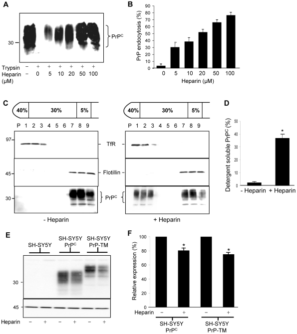
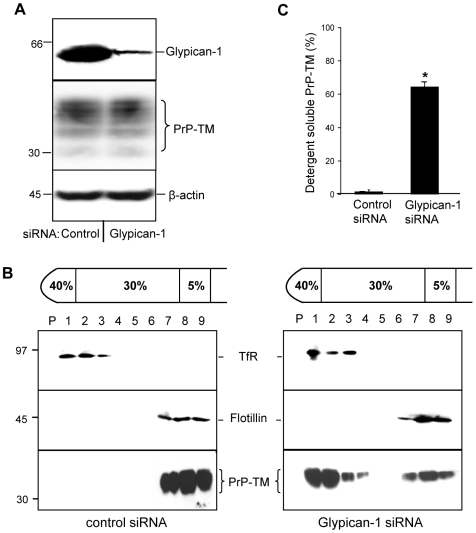
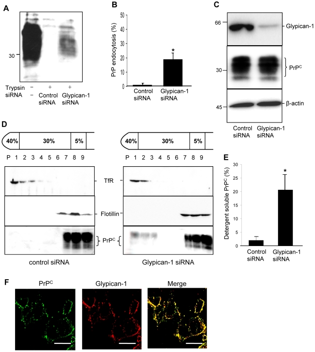
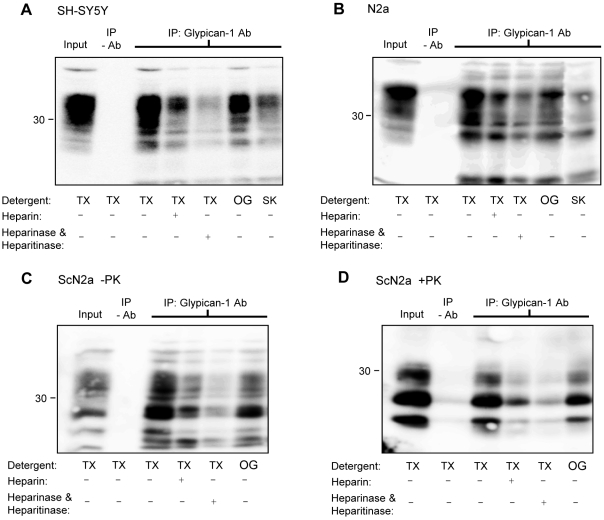
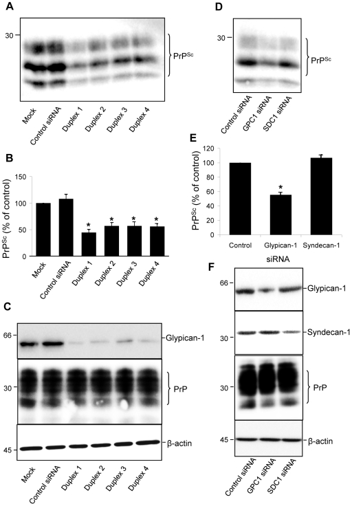
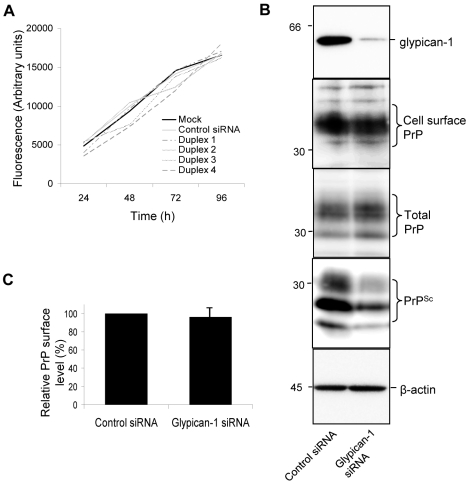
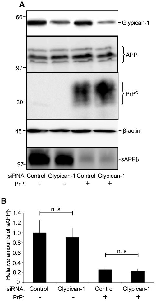
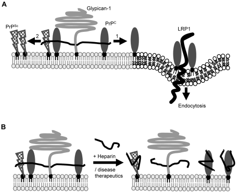
In prion diseases, the cellular form of the prion protein, PrP(C), undergoes a conformational conversion to the infectious isoform, PrP(Sc). PrP(C) associates with lipid rafts through its glycosyl-phosphatidylinositol (GPI) anchor and a region in its N-terminal domain which also binds to heparan sulfate proteoglycans (HSPGs). We show that heparin displaces PrP(C) from rafts and promotes its endocytosis, suggesting that heparin competes with an endogenous raft-resident HSPG for binding to PrP(C). We then utilised a transmembrane-anchored form of PrP (PrP-TM), which is targeted to rafts solely by its N-terminal domain, to show that both heparin and phosphatidylinositol-specific phospholipase C can inhibit its association with detergent-resistant rafts, implying that a GPI-anchored HSPG targets PrP(C) to rafts. Depletion of the major neuronal GPI-anchored HSPG, glypican-1, significantly reduced the raft association of PrP-TM and displaced PrP(C) from rafts, promoting its endocytosis. Glypican-1 and PrP(C) colocalised on the cell surface and both PrP(C) and PrP(Sc) co-immunoprecipitated with glypican-1. Critically, treatment of scrapie-infected N2a cells with glypican-1 siRNA significantly reduced PrP(Sc) formation. In contrast, depletion of glypican-1 did not alter the inhibitory effect of PrP(C) on the beta-secretase cleavage of the Alzheimer's amyloid precursor protein. These data indicate that glypican-1 is a novel cellular cofactor for prion conversion and we propose that it acts as a scaffold facilitating the interaction of PrP(C) and PrP(Sc) in lipid rafts.


Conflict of interest statement

The authors have declared that no competing interests exist.

Figures

Figure 1
Figure 1. Heparin stimulates the endocytosis of PrPC in a dose-dependent manner and displaces it from detergent-resistant lipid rafts.
(A) SH-SY5Y cells expressing PrPC were surface biotinylated and then incubated for 1 h at 37°C in the absence or presence of various concentrations of heparin diluted in OptiMEM. Prior to lysis cells were, where indicated, incubated with trypsin to digest cell surface PrPC. Cells were then lysed and PrPC immunoprecipitated from the sample using antibody 3F4. Samples were subjected to SDS PAGE and western blot analysis and the biotin-labelled PrPC detected with peroxidase-conjugated streptavidin. (B) Densitometric analysis of multiple blots from four separate experiments as described in (A) is shown. (C) SH-SY5Y cells expressing PrPC were surface biotinylated and then incubated in the absence or presence of 50 µM heparin prepared in OptiMEM for 1 h at 37°C. Cells were homogenised in the presence of 1% (v/v) Triton X-100 and subjected to buoyant sucrose density gradient centrifugation. PrPC was immunoprecipitated from equal volumes of each gradient fraction using 3F4 and subjected to SDS-PAGE and western blotting. The gradient fractions from both the untreated and heparin treated cells were analysed on the same SDS gel and immunoblotted under identical conditions. The biotin-labelled PrPC was detected with peroxidase-conjugated streptavidin. Flotillin-1 and transferrin receptor (TfR) were detected by immunoblotting as markers for DRM and detergent-soluble fractions, respectively. (D) Densitometric analysis of the proportion of total PrPC in the detergent soluble fractions of the plasma membrane. (E) Untransfected SH-SY5Y cells and SH-SY5Y cells expressing either PrPC or PrP-TM were grown to confluence and then incubated for 1 h in the presence or absence of 50 µM heparin prepared in OptiMEM. Media samples were collected and concentrated and cells harvested and lysed. Cell lysate samples were immunoblotted for PrPC using antibody 3F4, with β-actin used as a loading control. (F) Quantification of PrPC and PrP-TM levels after treatment of cells with heparin as in (E). Experiments were performed in triplicate and repeated on three occasions. * P<0.05.
Figure 2
Figure 2. The association of PrP-TM with DRMs is disrupted by treatment of cells with either heparin or bacterial PI-PLC.
SH-SY5Y cells expressing PrP-TM were surface biotinylated and then (A) incubated in the absence or presence of 50 µM heparin prepared in OptiMEM for 1 h at 37°C or (B) incubated in the absence or presence of 1 U/ml bacterial PI-PLC for 1 h at 4°C. Cells were homogenised in the presence of 1% (v/v) Triton X-100 and subjected to buoyant sucrose density gradient centrifugation. PrP-TM was immunoprecipitated from equal volumes of each gradient fraction using 3F4 and subjected to western blotting. The biotin-labelled PrP-TM fraction was detected with peroxidase-conjugated streptavidin. Flotillin-1 and transferrin receptor (TfR) were detected by immunoblotting as markers for DRM and detergent-soluble fractions respectively. (C) Densitometric analysis of the proportion of total PrP-TM present in the detergent soluble fractions of the plasma membrane after heparin and PI-PLC treatment. Experiments were performed in triplicate and repeated on three occasions. * P<0.05.
Figure 3
Figure 3. Depletion of glypican-1 inhibits the association of PrP-TM with DRMs.
SH-SY5Y cells expressing PrP-TM were treated with either control siRNA or siRNA targeted to glypican-1 and then allowed to reach confluence for 48 h. Cells were subsequently surface biotinylated and incubated in OptiMEM for 1 h at 37°C in the presence of Tyrphostin A23 to block endocytosis. The media was removed and the cells washed in phosphate-buffered saline prior to homogenisation in the presence of 1% (v/v) Triton X-100 and subjected to buoyant sucrose density gradient centrifugation. (A) Quantification of glypican-1 and PrP-TM expression in cell lysates. To detect glypican-1, cell lysate samples were treated with heparinase I and heparinase III prior to electrophoresis as described in the materials and methods section. (B) PrP-TM was immunoprecipitated from equal volumes of each gradient fraction using 3F4 and then subjected to western blotting with peroxidase-conjugated streptavidin. Flotillin-1 and transferrin receptor (TfR) were detected by immunoblotting as markers for DRM and detergent-soluble fractions, respectively. (C) Densitometric analysis of the proportion of total PrP-TM present in the detergent soluble fractions of the plasma membrane after siRNA treatment from multiple blots from three independent experiments. * P<0.05.
Figure 4
Figure 4. Depletion of glypican-1 stimulates the endocytosis of PrPC.
SH-SY5Y cells expressing wild type PrPC were treated with either control or glypican-1 siRNA and then incubated for 60 h. Cells were surface biotinylated and incubated in OptiMEM for 1 h at 37°C. Where indicated, cells were treated with trypsin to remove remaining cell surface PrPC. Cells were then lysed and total PrPC immunoprecipitated from the sample using antibody 3F4. (A) Samples were subjected to western blot analysis and the biotin-labelled PrPC fraction was detected with peroxidase-conjugated streptavidin. (B) Densitometric analysis (mean ± s.e.m.) of multiple blots from three separate experiments in (A) is shown. (C) Expression of glypican-1 (in lysate samples treated with heparinase I and heparinase III) and PrPC in the cell lysates from (A). β-actin was used as a loading control. (D) SH-SY5Y cells expressing PrPC were treated with either control siRNA or glypican-1 siRNA and then allowed to reach confluence for 48 h. Cells were subsequently surface biotinylated and incubated in OptiMEM for 1 h at 37°C. Cells were homogenised in the presence of 1% (v/v) Triton X-100 and subjected to buoyant sucrose density gradient centrifugation. (E) Densitometric analysis of the proportion of total PrPC present in the detergent soluble fractions of the plasma membrane after siRNA treatment from three independent experiments. (F) SH-SY5Y cells expressing PrPC were seeded onto glass coverslips and grown to 50% confluency. Cells were fixed, and then incubated with anti-PrP antibody 3F4 and a glypican-1 polyclonal antibody. Finally, cells were incubated with Alexa488-conjugated rabbit anti-mouse and Alexa594-conjugated goat anti-rabbit antibodies and viewed using a DeltaVision Optical Restoration Microscopy System. Images are representative of three individual experiments. Scale bars equal 10 µm. * P<0.05.
Figure 5
Figure 5. PrPC and PrPSc immunoprecipitate with glypican-1.
(A) SH-SY5Y cells expressing PrPC, (B) N2a cells or (C and D) ScN2a cells were lysed in the indicated detergents and then immunoprecipitated with a polyclonal glypican-1 antibody and where indicated, co-incubated with 50 µM heparin. Those samples pretreated with heparinase I and heparinase III were lysed with Triton X-100 followed by immunoprecipitation with glypican-1 antibody. In (D), immunopreciptiates from ScN2a cells were digested with PK. All immunoprecipitates were subsequently blotted for PrP. TX, Triton X-100; OG, octylglucoside; SK, sarkosyl.
Figure 6
Figure 6. Depletion of glypican-1 by siRNA reduces PrPSc formation.
ScN2a cells were either untreated or incubated with either control siRNA or one of four siRNAs targeted to glypican-1. After 48 h incubation the treatments were repeated. After a total incubation period of 96 h cells were harvested, lysed and protein concentration determined. (A) For detection of PK-resistant PrPSc, samples containing 200 µg protein were digested with 4 µg PK for 30 min at 37°C. Protein was then recovered by methanol precipitation and immunoblotted for PrP using antibody 6D11. (B) Densitometric analysis (mean ± s.e.m.) of PK-resistant PrPSc levels for each treatment, relative to those of mock-treated cells, from multiple blots from four independent experiments. (C) To confirm that glypican-1 depletion had been achieved in the ScN2a cells, cell lysate samples were immunoblotted for glypican-1, as well as PrP and β-actin. (D) To confirm the specificity of glypican-1 in modulating PrPSc formation, ScN2a cells were treated with control, syndecan-1 or glypican-1 siRNA reagents. Samples were processed as described in (A). (E) Densitometric analysis (mean ± s.e.m.) of PK-resistant PrPSc levels for each treatment in (D), from multiple blots from three independent experiments. (F) Confirmation of syndecan-1 and glypican-1 knockdown by immunblotting.
Figure 7
Figure 7. Depletion of glypican-1 does not affect cell division or surface levels of PrPC.
(A) ScN2a cells were seeded into 96 well plates and treated with transfection reagent only or incubated with either control siRNA or one of the four siRNAs targeted to glypican-1. Those experiments exceeding 48 h were dosed with a second treatment of the indicated siRNAs. Cells were then rinsed with PBS and fixed with 70% (v/v) ethanol. Plates were allowed to dry, stained with Hoescht 33342 and the fluorescence measured. (B) ScN2a cells were treated with control or glypican-1 siRNA. After 96 h, cell monolayers were labelled with a membrane impermeable biotin reagent. Biotin-labelled cell surface PrP was detected by immunoprecipitation using 6D11 and subsequent immunoblotting using HRP-conjugated streptavidin. Total PrP and PK-resistant PrP (PrPSc) were detected by immunoblotting using antibody 6D11. (C) Densitometric analysis of the proportion of the relative amount of biotinylated cell surface PrP in the absence or presence of glypican-1 siRNA from three independent experiments.
Figure 8
Figure 8. Depletion of glypican-1 does not affect the inhibition of BACE1 by PrPC.
SH-SY5Y cells co-expressing APP695 and PrP or cells expressing APP695 and an empty pIRESneo vector were treated with control siRNA or siRNA targeted to glypican-1. After 30 h the medium was replaced and cells incubated with reduced-serum medium for a further 24 h. Conditioned medium was harvested and cell lysates prepared. (A) Expression of glypican-1, full-length APP and PrPC in cell lysates, with β-actin as a loading control. Immunodetection of sAPPβ in conditioned medium. (B) Densitometric analysis of sAPPβ levels in glypican-1 depleted cells relative to control siRNA cells, calculated from multiple blots from three independent experiments.
Figure 9
Figure 9. Proposed model for glypican-1 in prion conversion.
Under normal circumstances, glypican-1 (light grey) with its heparan sulfate side chains (black lines) constitutes a lipid raft targeting determinant for PrPC. (A) For PrPC (dark grey) to exit lipid rafts and undergo endocytosis through its interaction with low density lipoprotein receptor-related protein 1 (LRP1, thick black line [59]), its interaction with glypican-1 must be disrupted (arrow 1). In prion disease, we hypothesise that glypican-1 provides a scaffold to facilitate interaction between PrPC and PrPSc (chequered) and allow misfolding of PrPC to proceed (arrow 2). (B) Prion therapeutic strategies may act, in part, by disrupting the interaction of PrPC and PrPSc that is facilitated through the heparan sulfate sidechains of glypican-1.

Similar articles

Cited by

References

    1. Prusiner SB. Prions. Proc Natl Acad Sci USA. 1998;95:13363–13383. - PMC - PubMed
    1. Taylor DR, Hooper NM. The prion protein and lipid rafts. Mol Membr Biol. 2006;23:89–99. - PubMed
    1. Hooper NM. Detergent-insoluble glycosphingolipid/cholesterol-rich membrane domains, lipid rafts and caveolae. Mol Membr Biol. 1999;16:145–156. - PubMed
    1. Baron GS, Caughey B. Effect of glycosylphosphatidylinositol anchor-dependent and -independent prion protein association with model raft membranes on conversion to the protease-resistant isoform. J Biol Chem. 2003;278:14883–14892. - PubMed
    1. Walmsley AR, Zeng F, Hooper NM. The N-terminal region of the prion protein ectodomain contains a lipid raft targeting determinant. J Biol Chem. 2003;278:37241–37248. - PubMed

Publication types