. 2009 Nov 19;4(11):e7897.
doi: 10.1371/journal.pone.0007897.

Catastrophic NAD+ depletion in activated T lymphocytes through Nampt inhibition reduces demyelination and disability in EAE

Affiliations

Catastrophic NAD+ depletion in activated T lymphocytes through Nampt inhibition reduces demyelination and disability in EAE

Santina Bruzzone et al. PLoS One. .

Abstract

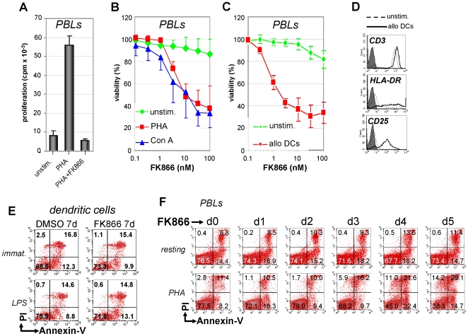
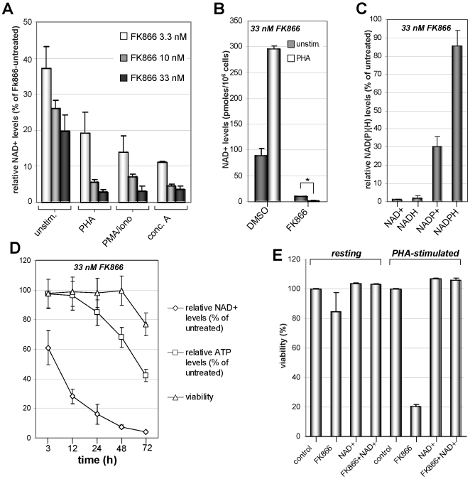
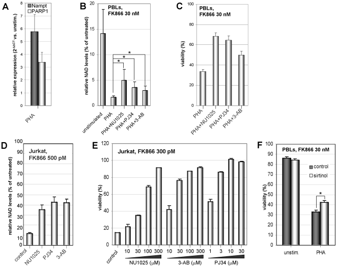
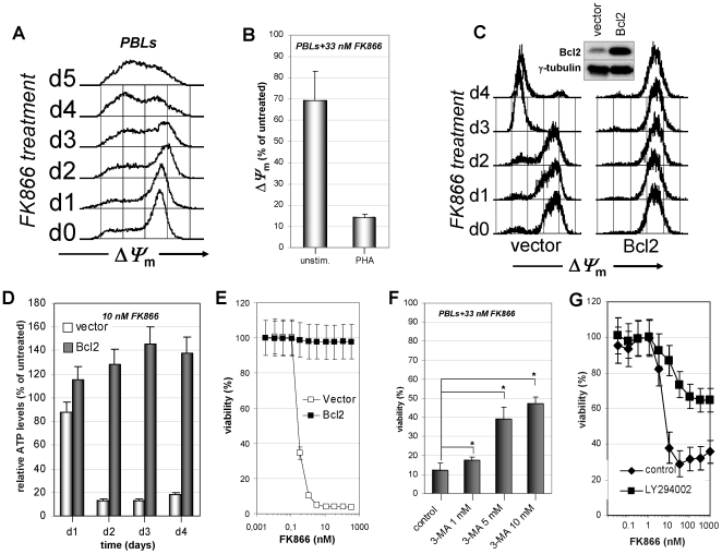
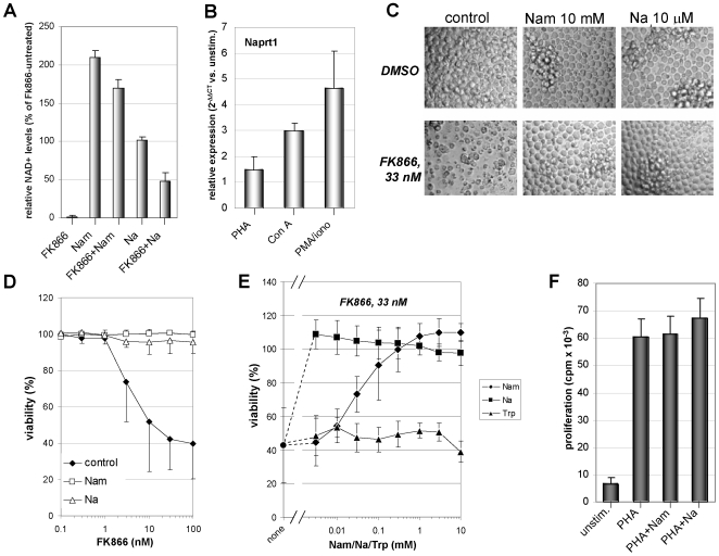
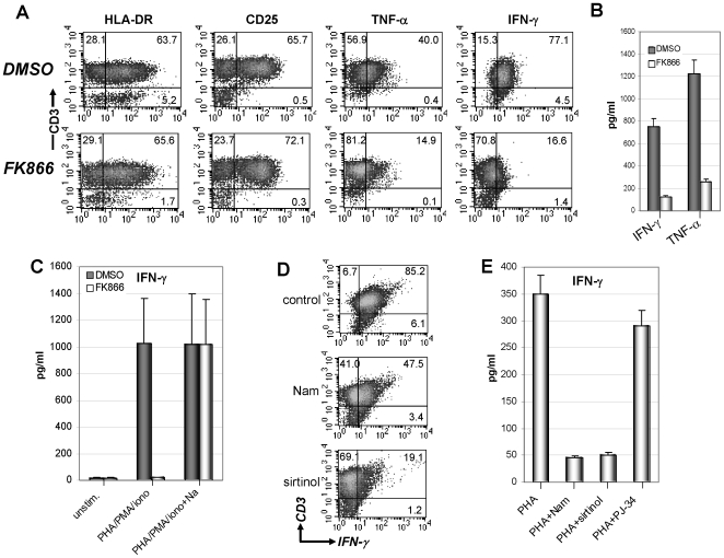
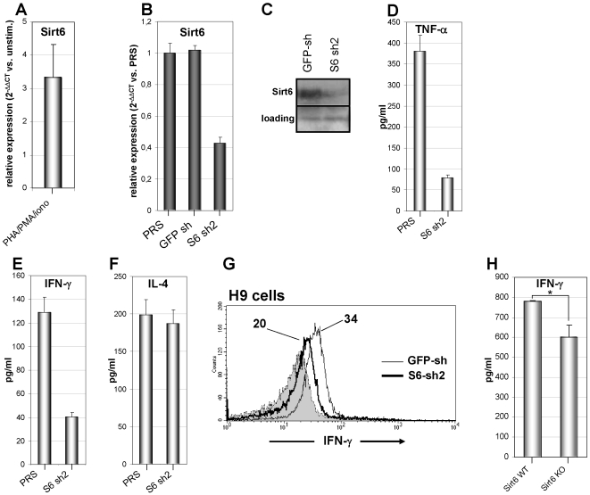
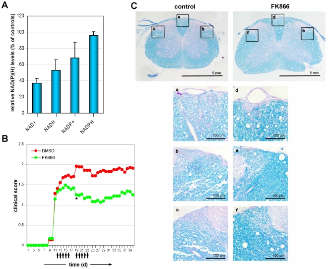
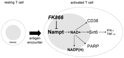
Nicotinamide phosphoribosyltransferase (Nampt) inhibitors such as FK866 are potent inhibitors of NAD(+) synthesis that show promise for the treatment of different forms of cancer. Based on Nampt upregulation in activated T lymphocytes and on preliminary reports of lymphopenia in FK866 treated patients, we have investigated FK866 for its capacity to interfere with T lymphocyte function and survival. Intracellular pyridine nucleotides, ATP, mitochondrial function, viability, proliferation, activation markers and cytokine secretion were assessed in resting and in activated human T lymphocytes. In addition, we used experimental autoimmune encephalomyelitis (EAE) as a model of T-cell mediated autoimmune disease to assess FK866 efficacy in vivo. We show that activated, but not resting, T lymphocytes undergo massive NAD(+) depletion upon FK866-mediated Nampt inhibition. As a consequence, impaired proliferation, reduced IFN-gamma and TNF-alpha production, and finally autophagic cell demise result. We demonstrate that upregulation of the NAD(+)-degrading enzyme poly-(ADP-ribose)-polymerase (PARP) by activated T cells enhances their susceptibility to NAD(+) depletion. In addition, we relate defective IFN-gamma and TNF-alpha production in response to FK866 to impaired Sirt6 activity. Finally, we show that FK866 strikingly reduces the neurological damage and the clinical manifestations of EAE. In conclusion, Nampt inhibitors (and possibly Sirt6 inhibitors) could be used to modulate T cell-mediated immune responses and thereby be beneficial in immune-mediated disorders.


Conflict of interest statement

Competing Interests: The authors have declared that no competing interests exist.

Figures

Figure 1
Figure 1. Nampt inhibition with FK866 prevents T lymphocyte proliferation and selectively kills activated T cells.
A, PBLs were seeded in 96-well plates in the presence or absence (unstim.) of PHA and 33 nM FK866. Proliferation was assessed 96 h later by standard [3H]thymidine incorporation assay. B, PBLs were incubated in 96-well plates in the presence or absence of 5 µg/ml PHA, 1 µg/ml Con A, with or without the indicated concentrations of FK866. Five days later viability was detected by PI staining and flow cytometry. Spontanous cell death was 12.2% and 28.1% for PHA- and Con A-stimulated PBLs, respectively. C, D, PBLs were stimulated for 7 days with or without allogeneic mature DCs before FK866 at the indicated concentrations was added. After 5 days viability was assessed by PI staining and flow cytometric analysis using the lymphocyte gate (C). Spontaneous PBL death was 18.4%. D: phenotype of unstimulated or DC-stimulated PBLs. E, Immature or LPS stimulated DCs were cultured for 7 days with 33 nM FK866 before staining with FITC-conjugated Annexin-V and PI and flow cytometry. F, Resting or PHA-stimulated PBLs were treated with 33 nM FK866 for the indicated times and subsequently stained with FITC-conjugated Annexin-V and PI for flow cytometric analysis. Mean values ± SD of five (B) and three (A, C) different donors are presented. D–F One representative experiment out of three is shown.
Figure 2
Figure 2. Activated T cells undergo massive NAD+ depletion upon Nampt inhibition.
A, 3×10⁶ PBLs/well were stimulated (or not, unstim.) with 5 µg/ml PHA, 1 µg/ml con A, or 50 ng/ml PMA and 0.5 µM ionomycin in the presence or absence of the indicated FK866 concentrations. 48 h later, cells were lysed in 0.6 M PCA and NAD+ content was measured in neutralized extracts. NAD+ levels were normalized to those detected in the absence of FK866. B, Unstimulated or PHA-stimulated PBLs were treated with 33 nM FK866 for 48 h before NAD+ content was determined. Absolute NAD+ levels are presented. *: p<0.05. C, PBLs were cultured for 48 h with PHA with or without FK866 (33 nM) addition. Subsequently, pyridine dinucleotides levels were measured in acid (NAD+ and NAPD+) or alkaline (NADH and NADPH) cell extracts. Dinucleotide levels were normalized to those detected without FK866. D, PBLs were incubated with PHA and 33 nM FK866 for the indicated times. Thereafter cells were harvested and NAD+ and ATP levels were determined in cell extracts whereas cell viability was assessed by PI-staining and flow cytometry. Results were normalized to the values of FK866-untreated cells. E, Resting or PHA-stimulated PBLs were treated (or not) with 33 nM FK866 in the presence or absence of 1 mM NAD+. After five-days viability was assessed determining PI cells by flow cytometry. Results are means ± SD of five (A) or three (B–E) experiments.
Figure 3
Figure 3. PARP inhibitors and sirtinol attenuate FK866-induced T cell demise.
A, PBLs were cultured for 24 h with or without PHA. Thereafter, Nampt and PARP1 levels were detected by Q-PCR. mRNA levels in PHA-stimulated cells were compared to those in unstimulated PBLs. B, Resting or PHA-stimulated PBLs were incubated with or without 33 nM FK866 in the presence or absence of 300 µM NU1025, 10 µM PJ34, or 300 µM 3-AB. 48 h later NAD+ levels were assessed (presented as % of values in FK866-untreated PBLs). *, p<0.05. C, PHA-stimulated PBLs were incubated for five days with or without 33 nM FK866 in the presence or absence of 300 µM NU1025, 10 µM PJ34, or 300 µM 3-AB. Thereafter, viability was assessed. D, 5×10⁵ Jurkat cells were treated for two days with 500 pM FK866 in the presence or absence of 300 µM NU1025, 5 µM PJ34, or 300 µM 3-AB. Subsequently, NAD+ content was determined and expressed as % of values in FK866-untreated Jurkat. E, 3×10⁴ Jurkat cells/well were incubated in 96-well plates with or without 300 pM FK866 in the presence or absence of the indicated concentrations of NU1025, PJ34, or 3-AB. Viability was determined 96 h later by PI cell staining and flow cytometry. F, PBLs were incubated for five days with PHA, with or without 33 nM FK866, in the presence or absence of 30 µM sirtinol. Viability was subsequently assessed by PI staining and flow cytometry. C, E, F, each treatment was tested in triplicate wells. Results are presented as means ± SD of three experiments.
Figure 4
Figure 4. Nampt inhibition with FK866 induces mitochondria depolarization and ATP depletion in activated T lymphocytes.
A, PHA- stimulated PBLs were incubated with 33 nM FK866 and ΔΨ m was determined at the indicated days of exposure. B, Resting of PHA-stimulated PBLs were cultured with 33 nM FK866 for five days. Thereafter PBLs with conserved ΔΨ m-high were quantified by flow-cytometry. C, Bcl2-overexpressing Jurkat and the respective vector control cells were incubated with 10 nM FK866 for the indicated number of days. Thereafter, ΔΨ m was determined. Inset, Western blot for Bcl2 and γ-tubulin expression. D, 5×10⁵ Bcl2-overexpressing Jurkat and the vector control cells were incubated with or without 10 nM FK866 for the indicated times before ATP was detected. ATP levels are presented as % of ATP in FK866-untreated cells. E, 3×10⁴ Bcl2-overexpressing and control Jurkat cells/well were incubated in 96-well plates with or without the indicated FK866 concentrations. Viability was determined by PI staining and flow cytometry 96 h later. F, PHA-stimulated PBLs were incubated in the presence or absence of 33 nM FK866 with or without the indicated concentrations of 3-MA. Viability was detected after five days. *, p<0.05. G, PBLs incubated in 96-well plates in the presence of 5 µg/ml PHA were treated for five days with the indicated FK866 concentrations in the presence or absence of 20 µM LY294002. Viability was subsequently determined by PI staining and flow cytometry. B, Results are presented as means ± SD of three experiments (B, D–G). Panels A and C are representative of three separate experiments.
Figure 5
Figure 5. Nam and Na prevent NAD+ shortage and cell death induced by FK866 in human T lymphocytes.
A, PHA-stimulated PBLs were treated (or not) with 33 nM FK866 in the presence or absence of 10 mM Nam or of 10 µM Na. After 48 h, NAD+ content was determined (expressed as percentage of NAD+ content in FK866-untreated cells). B, PBLs were cultured for 24 h with or without PHA, 1 µg/ml Con A, or 50 ng/ml PMA and 0.5 µM ionomycin. Thereafter, Naprt1 mRNA levels were detected by Q-PCR. mRNA levels in mitogen-stimulated PBLs were compared to those in unstimulated PBLs. C, D, PHA-stimulated PBLs were incubated for five days with or without 10 mM Nam or 10 µM Na in the presence or absence of the indicated FK866 concentrations. Thereafter, cells were imaged by light microscopy (C), and cell viability was determined (D). E, PHA-stimulated PBLs were incubated for five days with or without 33 nM Fk866 in the presence of the indicated concentrations of Nam, Na, or tryptophan (Trp). Viability was subsequently determined. F, PBLs were stimulated with or without PHA in the presence or absence of 1 mM Nam or Na. Thymidine incorporation was measured after 48 h by a 16-h pulse with 0.5 µCi/well [3H]thymidine. D-F each treatment was tested in triplicate wells. Results are means ± SD of three (A, B, F) or four (D, E) experiments. In panel C, one representative experiment out of three is shown.
Figure 6
Figure 6. Intracellular NAD+ depletion prevents TNF-α and IFN-γ production by activated T lymphocytes.
A, B, 5×10⁶ PBLs were stimulated with 5 µg/ml PHA with or without 33 nM FK866 for 36 h and subsequently incubated with 50 ng/ml PMA and 0.5 µM ionomycin for further 5 h. Thereafter, HLA-DR and CD25 expression were detected by flow cytometry. TNF-α and IFN-γ content was determined by intracellular cytokine staining and flow cytometry (A). Released TNF-α and IFN-γ were measured by ELISA (B). DMSO and FK866-treated cells were 84% and 83% viable, respectively. C, 5×10⁶ PBLs were stimulated with PHA and PMA/ionomycin as above, or left unstimulated. Where indicated, 33 nM FK866 with or without 10 µM Na was added. Released IFN-γ was measured by ELISA. Unstimulated PBLs treated with DMSO or FK866 were 96.3% and 96.5% viable, respectively. PBLs stimulated with PHA/PMA/ionomycin and treated with DMSO or FK866 were 89% and 89.5% viable, respectively. PBLs stimulated with PHA/PMA/ionomycin/Na and treated with DMSO or FK866 were 88% and 87% viable, respectively. D, PBLs were stimulated with PHA and PMA/ionomycin as describe for panel A. Where indicated 10 mM Nam or 50 µM sirtinol were added. Thereafter, cells were harvested and intracellular IFN-γ content was determined by flow cytometry. E, PBLs were stimulated with PHA and PMA/ionomycin as above in the presence or absence of 10 mM Nam, 50 µM sirtinol, or 10 µM PJ34. 42 h later, the supernatants were collected and IFN-γ levels were detected by ELISA. Results are means ± SD of four experiments (B, C, E). Panels A and D are representative of four separate experiments.
Figure 7
Figure 7. Evidence for an involvement of Sirt6 in IFN-γ synthesis.
A, PBLs were cultured for 24 h with or without PHA. Thereafter, Sirt6 levels were detected by Q-PCR. mRNA levels in PHA-stimulated cells were compared to those in unstimulated PBLs. B, C, Jurkat cells were transduced with PRS, (PRS) GFP-sh, or (PRS) S6 sh2, subsequently, Sirt6 mRNA levels or Sirt6 protein levels were determined by Q-PCR (B) and immunoblotting (C). D–F, Jurkat cells transduced with PRS or S6 sh2 were stimulated for 12 h with 5 µg/ml PHA, 50 ng/ml PMA, and 0.5 µM ionomycin. Thereafter, supernatants were harvested and TNF-α (D), IFN-γ (E), and IL-4 (F) levels were determined by ELISA. G, H9 cells transduced with GFP-sh or S6 sh2 were stimulated for 12 h with 5 µg/ml PHA, 50 ng/ml PMA, and 0.5 µM ionomycin. Thereafter, intracellular IFN-γ was detected by intracellular staining. Mean fluorescence intensity for IFN-γ expression is indicated for each histogram. H, 3×10⁶ splenocytes from wild type or Sirt6 KO mice/well were seeded in 24 well plates and stimulated for 24 h with 1 µg/ml Con A. Thereafter, supernatants were harvested and IFN-γ levels were determined by ELISA. *: p<0.05. Results are means ± SD of three experiments (A, B, D–F). Panel C and G are representative of three separate experiments.
Figure 8
Figure 8. FK866 ameliorates EAE.
10 mg/kg body weight FK866 were administered to mice from day 12 after rMOG immunization for 10 days. A, NAD(H) and NADP(H) levels were measured in mononuclear cells isolated from spleen and lymph nodes of treated or untreated animals at day 16. Dinucleotide levels in FK866-treated mice were expressed as % of those detected in control animals. B, FK866 halts EAE severity compared with controls (p<0.05 from day 19 onward). Arrows indicate the days of FK866 administration. C, Luxol fast Blue staining of the spinal cord shows areas of demyelination in control mice compared with FK866-treated animals.
Figure 9
Figure 9. A putative model of Nampt's role in activated T lymphocytes.
Nampt activity is responsible for providing sufficient NAD+ supplies during T cell activation. NAD+, in turn, is required for ATP synthesis, metabolic reactions, and to replenish NADPH levels. In addition, NAD+ represents the substrate of NAD+-degrading enzymes such as PARP, CD38, and the sirtuins. Among these, Sirt6 appears to have a central role in IFN-γ and TNF-α production. Nampt inhibitors such as FK866 (and possibly Sirt6 inhibitors) could be used to modulate T cell-mediated immune responses and thereby be beneficial in immune disorders.

Similar articles

Cited by

References

    1. Hasmann M, Schemainda I. FK866, a highly specific noncompetitive inhibitor of nicotinamide phosphoribosyltransferase, represents a novel mechanism for induction of tumor cell apoptosis. Cancer Res. 2003;63:7436–7442. - PubMed
    1. Khan JA, Tao X, Tong L. Molecular basis for the inhibition of human NMPRTase, a novel target for anticancer agents. Nat Struct Mol Biol. 2006;13:582–588. - PubMed
    1. Wosikowski K, Mattern K, Schemainda I, Hasmann M, Rattel B, et al. WK175, a novel antitumor agent, decreases the intracellular nicotinamide adenine dinucleotide concentration and induces the apoptotic cascade in human leukemia cells. Cancer Res. 2002;62:1057–1062. - PubMed
    1. Nahimana A, Attinger A, Aubry D, Greaney P, Ireson C, et al. The NAD biosynthesis inhibitor APO866 has potent antitumor activity against hematologic malignancies. Blood. 2009;113:3276–3286. - PubMed
    1. Holen K, Saltz LB, Hollywood E, Burk K, Hanauske AR. The pharmacokinetics, toxicities, and biologic effects of FK866, a nicotinamide adenine dinucleotide biosynthesis inhibitor. Invest New Drugs. 2008;26:45–51. - PubMed

Publication types

MeSH terms