

Role of fetal stem cells in maternal tissue regeneration
- PMID: 19936082
- PMCID: PMC2759120
Role of fetal stem cells in maternal tissue regeneration
Abstract
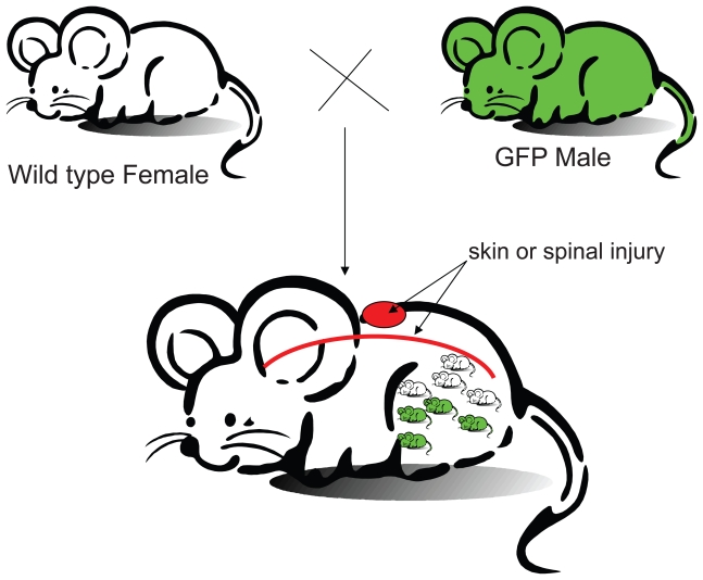
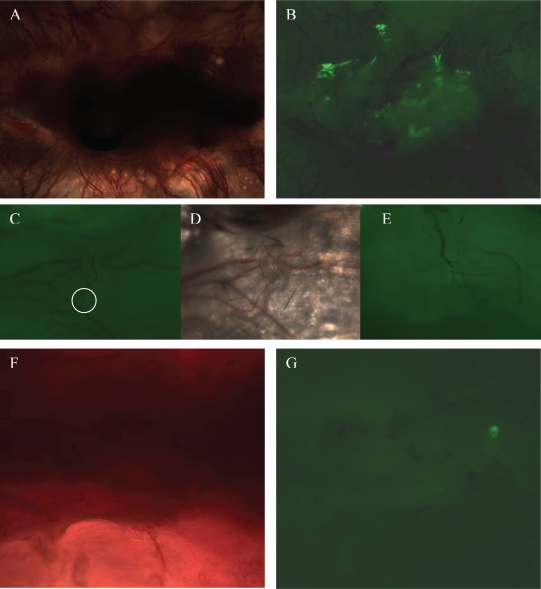
Microchimerism refers to the status of harboring cells from another individual at low levels. It is well known that cells traffic bidirectionally between fetus and mother during pregnancy. This situation resembles a naturally occurring long lasting fetal stem cell transplantation. The fetus acts as the donor and the mother acts as the recipient. To study the role of microchimerism in tissue regeneration, we constructed a murine microchimerism model with wild type C57BL/6J female mice carrying progenies which expressed green fluorescent proteins (GFP). Our data indicated that skin injuries in the female mice during pregnancy increased microchimerism of GFP expressing cells from the GFP transgenic progenies. The GFP positive cells also appeared at the site of spinal cord where injury occurred during pregnancy. Our study suggests that the amount of fetal cells in maternal mice significantly increased if injuries occurred during pregnancy. Fetal stem cells appear to respond to maternal injury signals and may play a role in maternal tissue regeneration during pregnancy.
Keywords: Fetal stem cells; Injury introduction; Microchimerism.
Figures
Similar articles
-
Fetal cell microchimerism in the maternal mouse spinal cord.Neurosci Bull. 2014 Feb;30(1):81-9. doi: 10.1007/s12264-013-1392-1. Epub 2013 Dec 17. Neurosci Bull. 2014. PMID: 24346789 Free PMC article.
-
Long-term feto-maternal microchimerism: nature's hidden clue for alternative donor hematopoietic cell transplantation?Int J Hematol. 2002 Oct;76(3):229-37. doi: 10.1007/BF02982792. Int J Hematol. 2002. PMID: 12416733
-
Natural history of fetal cell microchimerism during and following murine pregnancy.J Reprod Immunol. 2005 Jun;66(1):1-12. doi: 10.1016/j.jri.2005.02.001. J Reprod Immunol. 2005. PMID: 15949558
-
Fetal microchimerism: benevolence or malevolence for the mother?Eur J Obstet Gynecol Reprod Biol. 2011 Oct;158(2):148-52. doi: 10.1016/j.ejogrb.2011.05.008. Epub 2011 Jun 12. Eur J Obstet Gynecol Reprod Biol. 2011. PMID: 21664033 Review.
-
Fetal-maternal microchimerism: impact on hematopoietic stem cell transplantation.Curr Opin Immunol. 2005 Oct;17(5):546-52. doi: 10.1016/j.coi.2005.07.009. Curr Opin Immunol. 2005. PMID: 16084712 Review.
Cited by
-
Pregnancy-associated progenitor cells: an under-recognized potential source of stem cells in maternal lung.Placenta. 2011 Oct;32 Suppl 4:S298-303. doi: 10.1016/j.placenta.2011.04.007. Epub 2011 May 4. Placenta. 2011. PMID: 21546085 Free PMC article. Review.
-
Twisting immune responses for allogeneic stem cell therapy.World J Stem Cells. 2009 Dec 31;1(1):30-5. doi: 10.4252/wjsc.v1.i1.30. World J Stem Cells. 2009. PMID: 20975985 Free PMC article.
References
-
- Adams KM, Nelson JL. Microchimerism: an investigative frontier in autoimmunity and transplantation. JAMA. 2004;291(9):1127–31. - PubMed
-
- Aractingi S, Khosrotehrani K. Microchimerism: fears and hopes.[comment] Dermatology. 2005;210(1):1–2. - PubMed
-
- Artlett CM, Smith JB, et al. Identification of fetal DNA and cells in skin lesions from women with systemic sclerosis.[see comment] N Engl J Med. 1998;338(17):1186–91. - PubMed
-
- Bianchi DW, Farina A, et al. Significant fetal-maternal hemorrhage after termination of pregnancy: implications for development of fetal cell microchimerism. American Journal of Obstetrics & Gynecology. 2001;184(4):703–6. - PubMed
LinkOut - more resources
Full Text Sources