

Morphological and molecular changes of the myocardium after left ventricular mechanical support
- PMID: 19936192
- PMCID: PMC2780817
- DOI: 10.2174/157340308785160606
Morphological and molecular changes of the myocardium after left ventricular mechanical support
Abstract
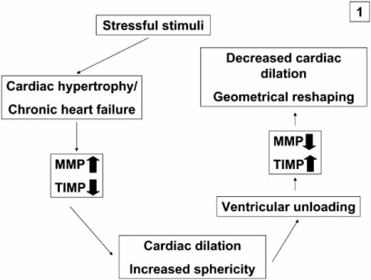
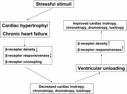
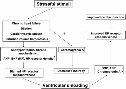
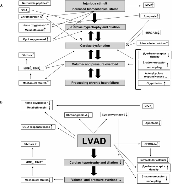
Left ventricular assist devices (LVAD) are currently used to either "bridge" patients with terminal congestive heart failure (CHF) until cardiac transplantation is possible or optionally for patients with contraindications for transplantation ("destination therapy"). Mechanical support is associated with a marked decrease of cardiac dilation and hypertrophy as well as numerous cellular and molecular changes ("reverse cardiac remodeling"), which can be accompanied by improved cardiac function ("bridge to recovery") in a relatively small subset of patients with heart transplantation no longer necessary even after removal of the device ("weaning"). In the recent past, novel pharmacological strategies have been developed and are combined with mechanical support, which has increased the percentage of patients with improved clinical status and cardiac performance. Gene expression profiles have demonstrated that individuals who recover after LVAD show different gene expression compared to individuals who do not respond to unloading. This methodology holds promise for the future to develop read out frames to identify individuals who can recover after support. Aside from describing the morphological changes associated with "reverse cardiac remodeling", this review will focus on signal transduction, transcriptional regulation, apoptosis, cell stress proteins, matrix remodeling, inflammatory mediators and aspects of neurohormonal activation in the failing human heart before and after ventricular unloading.
Keywords: Congestive heart failure (CHF); left ventricular assist device (LVAD); morphology; reverse cardiac remodeling; ventricular unloading; weaning..
Figures
Similar articles
-
[Congestive heart failure: reverse cardiac remodeling mediated by left ventricular assist devices].Pathologe. 2012 May;33(3):175-82. doi: 10.1007/s00292-011-1559-3. Pathologe. 2012. PMID: 22576594 German.
-
Reverse remodeling following insertion of left ventricular assist devices (LVAD): a review of the morphological and molecular changes.Cardiovasc Res. 2005 Dec 1;68(3):376-86. doi: 10.1016/j.cardiores.2005.06.030. Epub 2005 Jul 18. Cardiovasc Res. 2005. PMID: 16024006 Review.
-
Sarcomeric genes involved in reverse remodeling of the heart during left ventricular assist device support.J Heart Lung Transplant. 2005 Jan;24(1):73-80. doi: 10.1016/j.healun.2003.10.016. J Heart Lung Transplant. 2005. PMID: 15653383
-
Molecular changes after left ventricular assist device support for heart failure.Circ Res. 2013 Aug 30;113(6):777-91. doi: 10.1161/CIRCRESAHA.113.301413. Circ Res. 2013. PMID: 23989719 Review.
-
[Left ventricular assist devices (LVAD): optional treatment alternative to cardiac transplantation?].Verh Dtsch Ges Pathol. 2004;88:113-21. Verh Dtsch Ges Pathol. 2004. PMID: 16892541 Review. German.
Cited by
-
[Congestive heart failure: reverse cardiac remodeling mediated by left ventricular assist devices].Pathologe. 2012 May;33(3):175-82. doi: 10.1007/s00292-011-1559-3. Pathologe. 2012. PMID: 22576594 German.
-
Bridge to recovery: understanding the disconnect between clinical and biological outcomes.Circulation. 2012 Jul 10;126(2):230-41. doi: 10.1161/CIRCULATIONAHA.111.040261. Circulation. 2012. PMID: 22777666 Free PMC article. No abstract available.
-
Modeling of Isoproterenol-Induced Chronic Heart Failure in 24-Month-Old Rats.Bull Exp Biol Med. 2024 Nov;178(1):30-33. doi: 10.1007/s10517-024-06277-8. Epub 2024 Nov 22. Bull Exp Biol Med. 2024. PMID: 39572489
-
Temporal pattern of left ventricular structural and functional remodeling following reversal of volume overload heart failure.J Appl Physiol (1985). 2011 Dec;111(6):1778-88. doi: 10.1152/japplphysiol.00691.2011. Epub 2011 Sep 1. J Appl Physiol (1985). 2011. PMID: 21885799 Free PMC article.
-
An Improved UNet++ Model for Congestive Heart Failure Diagnosis Using Short-Term RR Intervals.Diagnostics (Basel). 2021 Mar 16;11(3):534. doi: 10.3390/diagnostics11030534. Diagnostics (Basel). 2021. PMID: 33809773 Free PMC article.
References
-
- Boucek MM, Edwards LB, Keck BM, et al. Registry for the International Society for Heart and Lung Transplantation: seventh official pediatric report-2004. J Heart Lung Transplant. 2004;23:933–947. - PubMed
-
- Taylor DO, Edwards LB, Boucek MM, et al. The Registry of the International Society for Heart and Lung Transplantation: twenty-first official adult heart transplant report 2004. J Heart Lung Transplant. 2004;23:796–803. - PubMed
-
- Sliwa K, Damasceno A, Mayosi BM. Epidemiology and etiology of cardiomyopathy in Africa. Circulation. 2005;112:3577–3583. - PubMed
-
- Goldstein DJ, Oz MC, Rose EA. Implantable left ventricular assist devices. N Engl J Med. 1998;339:1522–1533. - PubMed
-
- Barbone A, Holmes JW, Heerdt PM, et al. Comparison of right and left ventricular responses to left ventricular assist device support in patients with severe heart failure: a primary role of mechanical unloading underlying reverse remodeling. Circulation. 2001;104:670–675. - PubMed
LinkOut - more resources
Full Text Sources
Miscellaneous
