

Disorganized innervation and neuronal loss in the inner ear of Slitrk6-deficient mice
- PMID: 19936227
- PMCID: PMC2777407
- DOI: 10.1371/journal.pone.0007786
Disorganized innervation and neuronal loss in the inner ear of Slitrk6-deficient mice
Abstract
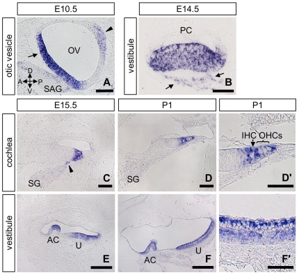
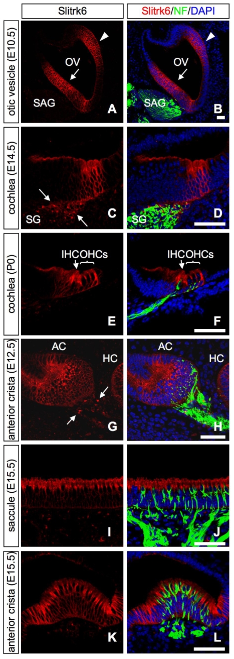
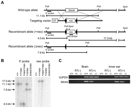
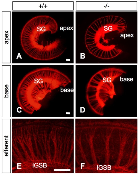
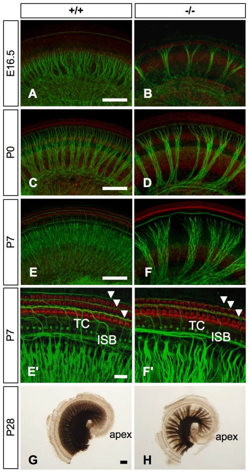
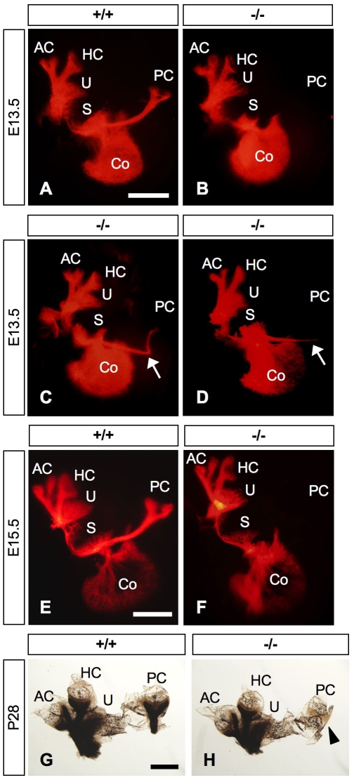
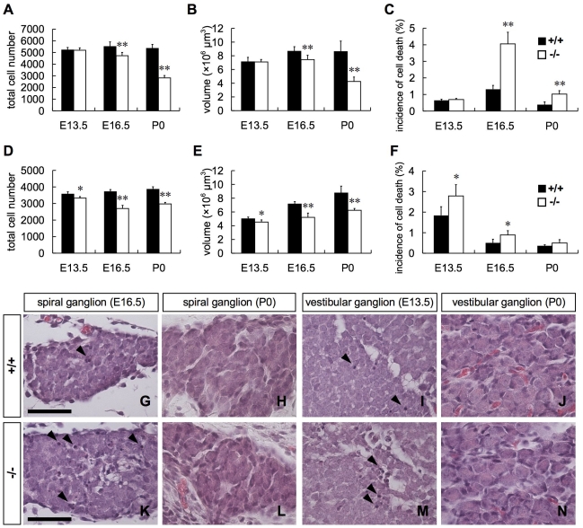
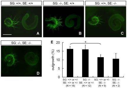
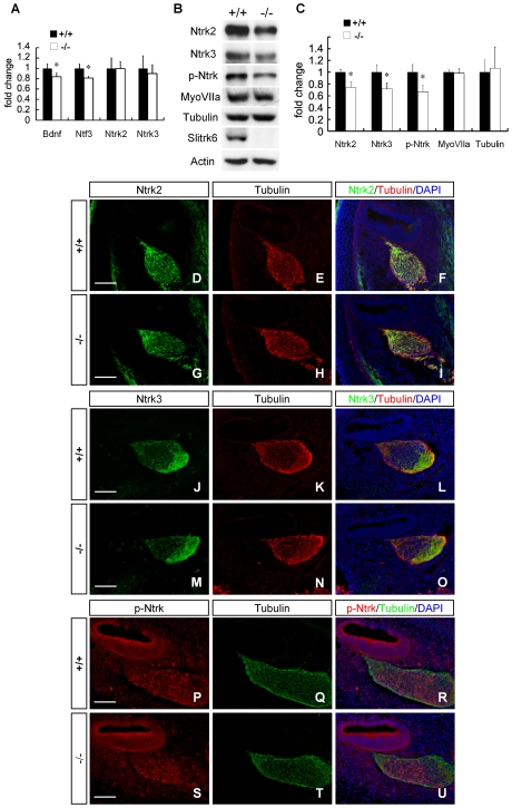
Slitrks are type I transmembrane proteins that share conserved leucine-rich repeat domains similar to those in the secreted axonal guidance molecule Slit. They also show similarities to Ntrk neurotrophin receptors in their carboxy-termini, sharing a conserved tyrosine residue. Among 6 Slitrk family genes in mammals, Slitrk6 has a unique expression pattern, with strong expression in the sensory epithelia of the inner ear. We generated Slitrk6-knockout mice and investigated the development of their auditory and vestibular sensory organs. Slitrk6-deficient mice showed pronounced reduction in the cochlear innervation. In the vestibule, the innervation to the posterior crista was often lost, reduced, or sometimes misguided. These defects were accompanied by the loss of neurons in the spiral and vestibular ganglia. Cochlear sensory epithelia from Slitrk6-knockout mice have reduced ability in promoting neurite outgrowth of spiral ganglion neurons. Indeed the Slitrk6-deficient inner ear showed a mild but significant decrease in the expression of Bdnf and Ntf3, both of which are essential for the innervation and survival of sensory neurons. In addition, the expression of Ntrk receptors, including their phosphorylated forms was decreased in Slitrk6-knockout cochlea. These results suggest that Slitrk6 promotes innervation and survival of inner ear sensory neurons by regulating the expression of trophic and/or tropic factors including neurotrophins from sensory epithelia.
Conflict of interest statement
Figures
Similar articles
-
Neurod1 regulates survival and formation of connections in mouse ear and brain.Cell Tissue Res. 2010 Jul;341(1):95-110. doi: 10.1007/s00441-010-0984-6. Epub 2010 May 30. Cell Tissue Res. 2010. PMID: 20512592 Free PMC article.
-
Brn3c null mutant mice show long-term, incomplete retention of some afferent inner ear innervation.BMC Neurosci. 2003 Jan 30;4:2. doi: 10.1186/1471-2202-4-2. BMC Neurosci. 2003. PMID: 12585968 Free PMC article.
-
Mutant mice reveal the molecular and cellular basis for specific sensory connections to inner ear epithelia and primary nuclei of the brain.Hear Res. 2005 Aug;206(1-2):52-63. doi: 10.1016/j.heares.2004.11.025. Hear Res. 2005. PMID: 16080998 Free PMC article. Review.
-
Neurotrophins in the ear: their roles in sensory neuron survival and fiber guidance.Prog Brain Res. 2004;146:265-78. doi: 10.1016/S0079-6123(03)46017-2. Prog Brain Res. 2004. PMID: 14699969 Review.
-
Coordinated expression and function of neurotrophins and their receptors in the rat inner ear during target innervation.Hear Res. 1994 May;75(1-2):131-44. doi: 10.1016/0378-5955(94)90064-7. Hear Res. 1994. PMID: 8071140
Cited by
-
Neurod1 regulates survival and formation of connections in mouse ear and brain.Cell Tissue Res. 2010 Jul;341(1):95-110. doi: 10.1007/s00441-010-0984-6. Epub 2010 May 30. Cell Tissue Res. 2010. PMID: 20512592 Free PMC article.
-
Lineage-tracing and translatomic analysis of damage-inducible mitotic cochlear progenitors identifies candidate genes regulating regeneration.PLoS Biol. 2021 Nov 10;19(11):e3001445. doi: 10.1371/journal.pbio.3001445. eCollection 2021 Nov. PLoS Biol. 2021. PMID: 34758021 Free PMC article.
-
Pegylated Insulin-Like Growth Factor 1 attenuates Hair Cell Loss and promotes Presynaptic Maintenance of Medial Olivocochlear Cholinergic Fibers in the Cochlea of the Progressive Motor Neuropathy Mouse.Front Neurol. 2022 Jun 3;13:885026. doi: 10.3389/fneur.2022.885026. eCollection 2022. Front Neurol. 2022. PMID: 35720065 Free PMC article.
-
GATA3 maintains the quiescent state of cochlear supporting cells by regulating p27kip1.Sci Rep. 2021 Aug 4;11(1):15779. doi: 10.1038/s41598-021-95427-3. Sci Rep. 2021. PMID: 34349220 Free PMC article.
-
Slitrk4 is required for the development of inhibitory neurons in the fear memory circuit of the lateral amygdala.Front Mol Neurosci. 2024 Apr 26;17:1386924. doi: 10.3389/fnmol.2024.1386924. eCollection 2024. Front Mol Neurosci. 2024. PMID: 38736483 Free PMC article.
References
-
- Aruga J, Mikoshiba K. Identification and characterization of Slitrk, a novel neuronal transmembrane protein family controlling neurite outgrowth. Mol Cell Neurosci. 2003;24:117–129. - PubMed
-
- Aruga J, Yokota N, Mikoshiba K. Human SLITRK family genes: genomic organization and expression profiling in normal brain and brain tumor tissue. Gene. 2003;315:87–94. - PubMed
-
- Kobe B, Kajava AV. The leucine-rich repeat as a protein recognition motif. Curr Opin Struct Biol. 2001;11:725–732. - PubMed
-
- Brose K, Tessier-Lavigne M. Slit proteins: key regulators of axon guidance, axonal branching, and cell migration. Curr Opin Neurobiol. 2000;10:95–102. - PubMed
-
- Huang EJ, Reichardt LF. Trk receptors: roles in neuronal signal transduction. Annu Rev Biochem. 2003;72:609–642. - PubMed
