

Administration of Mycobacterium leprae rHsp65 aggravates experimental autoimmune uveitis in mice
- PMID: 19936251
- PMCID: PMC2775913
- DOI: 10.1371/journal.pone.0007912
Administration of Mycobacterium leprae rHsp65 aggravates experimental autoimmune uveitis in mice
Abstract
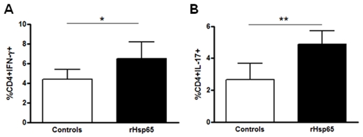
The 60 kDa heat shock protein family, Hsp60, constitutes an abundant and highly conserved class of molecules that are highly expressed in chronic-inflammatory and autoimmune processes. Experimental autoimmune uveitis [EAU] is a T cell mediated intraocular inflammatory disease that resembles human uveitis. Mycobacterial and homologous Hsp60 peptides induces uveitis in rats, however their participation in aggravating the disease is poorly known. We here evaluate the effects of the Mycobacterium leprae Hsp65 in the development/progression of EAU and the autoimmune response against the eye through the induction of the endogenous disequilibrium by enhancing the entropy of the immunobiological system with the addition of homologous Hsp. B10.RIII mice were immunized subcutaneously with interphotoreceptor retinoid-binding protein [IRBP], followed by intraperitoneally inoculation of M. leprae recombinant Hsp65 [rHsp65]. We evaluated the proliferative response, cytokine production and the percentage of CD4(+)IL-17(+), CD4(+)IFN-gamma(+) and CD4(+)Foxp3(+) cells ex vivo, by flow cytometry. Disease severity was determined by eye histological examination and serum levels of anti-IRBP and anti-Hsp60/65 measured by ELISA. EAU scores increased in the Hsp65 group and were associated with an expansion of CD4(+)IFN-gamma(+) and CD4(+)IL-17(+) T cells, corroborating with higher levels of IFN-gamma. Our data indicate that rHsp65 is one of the managers with a significant impact over the immune response during autoimmunity, skewing it to a pathogenic state, promoting both Th1 and Th17 commitment. It seems comprehensible that the specificity and primary function of Hsp60 molecules can be considered as a potential pathogenic factor acting as a whistleblower announcing chronic-inflammatory diseases progression.
Conflict of interest statement
Figures


Similar articles
-
SLAT/Def6 plays a critical role in the pathogenic process of experimental autoimmune uveitis (EAU).Mol Vis. 2012;18:1858-64. Epub 2012 Jul 7. Mol Vis. 2012. PMID: 22815639 Free PMC article.
-
Tofacitinib inhibits the development of experimental autoimmune uveitis and reduces the proportions of Th1 but not of Th17 cells.Mol Vis. 2020 Sep 26;26:641-651. eCollection 2020. Mol Vis. 2020. PMID: 33088168 Free PMC article.
-
CTLA4-Ig suppresses development of experimental autoimmune uveitis in the induction and effector phases: Comparison with blockade of interleukin-6.Exp Eye Res. 2015 Nov;140:53-64. doi: 10.1016/j.exer.2015.08.012. Epub 2015 Aug 20. Exp Eye Res. 2015. PMID: 26297802
-
The immunopathogenesis of chronic and relapsing autoimmune uveitis - Lessons from experimental rat models.Prog Retin Eye Res. 2018 Jul;65:107-126. doi: 10.1016/j.preteyeres.2018.02.003. Epub 2018 Feb 27. Prog Retin Eye Res. 2018. PMID: 29496590 Review.
-
Role of CD4+ T cell-derived cytokines in the pathogenesis of uveitis.Clin Exp Med. 2025 Feb 5;25(1):49. doi: 10.1007/s10238-025-01565-7. Clin Exp Med. 2025. PMID: 39909966 Free PMC article. Review.
Cited by
-
A Mycobacterium leprae Hsp65 mutant as a candidate for mitigating lupus aggravation in mice.PLoS One. 2011;6(9):e24093. doi: 10.1371/journal.pone.0024093. Epub 2011 Sep 22. PLoS One. 2011. PMID: 21961033 Free PMC article.
-
Mycobacterium leprae Hsp65 administration reduces the lifespan of aged high antibody producer mice.Immun Ageing. 2014 Mar 26;11(1):6. doi: 10.1186/1742-4933-11-6. Immun Ageing. 2014. PMID: 24669842 Free PMC article.
-
Novel CD28 antagonist mPEG PV1-Fab' mitigates experimental autoimmune uveitis by suppressing CD4+ T lymphocyte activation and IFN-γ production.PLoS One. 2017 Mar 1;12(3):e0171822. doi: 10.1371/journal.pone.0171822. eCollection 2017. PLoS One. 2017. PMID: 28248972 Free PMC article.
-
Immunopathophysiology and clinical impact of uveitis in inflammatory rheumatic diseases: An update.Eur J Clin Invest. 2021 Aug;51(8):e13572. doi: 10.1111/eci.13572. Epub 2021 May 5. Eur J Clin Invest. 2021. PMID: 33851422 Free PMC article. Review.
-
HSP60: a double edge sword in autoimmunity.Oncotarget. 2015 Oct 20;6(32):32299-300. doi: 10.18632/oncotarget.5869. Oncotarget. 2015. PMID: 26431161 Free PMC article. No abstract available.
References
-
- Whitcup SM, Nussenblatt RB. Immunologic mechanisms of uveitis. New targets for immunomodulation. Arch Ophthalmol. 1997;115:520–525. - PubMed
-
- Caspi RR, Roberge FG, Chan CC, Wiggert B, Chader GJ, et al. A new model of autoimmune disease. Experimental autoimmune uveoretinitis induced in mice with two different retinal antigens. J Immunol. 1988;140:1490–1495. - PubMed
-
- Mochizuki M, Kuwabara T, McAllister C, Nussenblatt RB, Gery I. Adoptive transfer of experimental autoimmune uveoretinitis in rats. Immunopathogenic mechanisms and histologic features. Invest Ophthalmol Vis Sci. 1985;26:1–9. - PubMed
-
- Rizzo LV, Silver P, Wiggert B, Hakim F, Gazzinelli RT, et al. Establishment and characterization of a murine CD4+ T cell line and clone that induce experimental autoimmune uveoretinitis in B10.A mice. J Immunol. 1996;156:1654–1660. - PubMed
-
- Thole JE, Hindersson P, de Bruyn J, Cremers F, van der Zee J, et al. Antigenic relatedness of a strongly immunogenic 65 kDA mycobacterial protein antigen with a similarly sized ubiquitous bacterial common antigen. Microb Pathog. 1988;4:71–83. - PubMed
Publication types
MeSH terms
Substances
LinkOut - more resources
Full Text Sources
Medical
Research Materials
Miscellaneous
