. 2009 Nov 20;4(11):e7930.
doi: 10.1371/journal.pone.0007930.

Lrp4, a novel receptor for Dickkopf 1 and sclerostin, is expressed by osteoblasts and regulates bone growth and turnover in vivo

Affiliations

Lrp4, a novel receptor for Dickkopf 1 and sclerostin, is expressed by osteoblasts and regulates bone growth and turnover in vivo

Hong Y Choi et al. PLoS One. .

Abstract

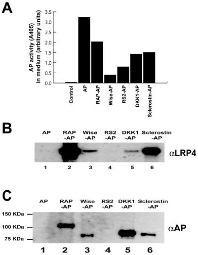
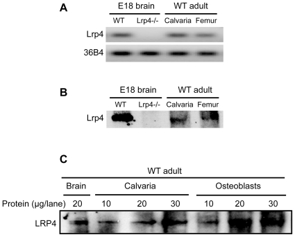
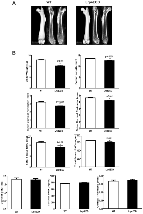
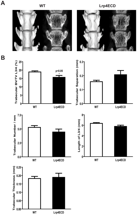
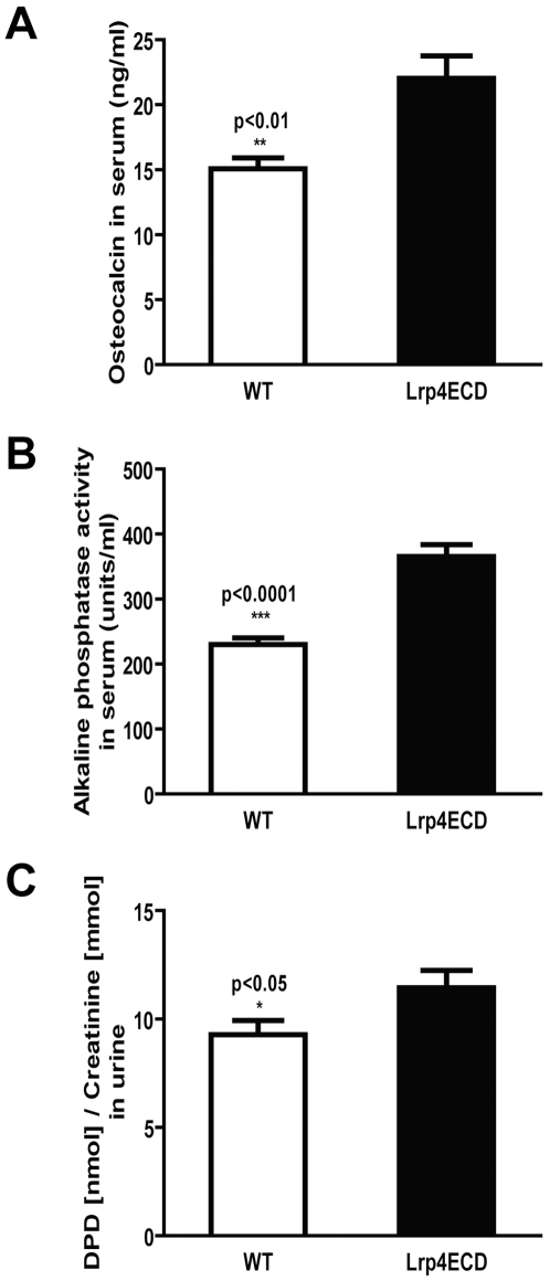
Lrp4 is a multifunctional member of the low density lipoprotein-receptor gene family and a modulator of extracellular cell signaling pathways in development. For example, Lrp4 binds Wise, a secreted Wnt modulator and BMP antagonist. Lrp4 shares structural elements within the extracellular ligand binding domain with Lrp5 and Lrp6, two established Wnt co-receptors with important roles in osteogenesis. Sclerostin is a potent osteocyte secreted inhibitor of bone formation that directly binds Lrp5 and Lrp6 and modulates both BMP and Wnt signaling. The anti-osteogenic effect of sclerostin is thought to be mediated mainly by inhibition of Wnt signaling through Lrp5/6 within osteoblasts. Dickkopf1 (Dkk1) is another potent soluble Wnt inhibitor that binds to Lrp5 and Lrp6, can displace Lrp5-bound sclerostin and is itself regulated by BMPs. In a recent genome-wide association study of bone mineral density a significant modifier locus was detected near the SOST gene at 17q21, which encodes sclerostin. In addition, nonsynonymous SNPs in the LRP4 gene were suggestively associated with bone mineral density. Here we show that Lrp4 is expressed in bone and cultured osteoblasts and binds Dkk1 and sclerostin in vitro. MicroCT analysis of Lrp4 deficient mutant mice revealed shortened total femur length, reduced cortical femoral perimeter, and reduced total femur bone mineral content (BMC) and bone mineral density (BMD). Lumbar spine trabecular bone volume per total volume (BV/TV) was significantly reduced in the mutants and the serum and urinary bone turnover markers alkaline phosphatase, osteocalcin and desoxypyridinoline were increased. We conclude that Lrp4 is a novel osteoblast expressed Dkk1 and sclerostin receptor with a physiological role in the regulation of bone growth and turnover, which is likely mediated through its function as an integrator of Wnt and BMP signaling pathways.


Conflict of interest statement

Competing Interests: A.N. received a short-term travel grant from Merck, Sharpe and Dohme. This has or had no influence on data collection, experimental planning, data interpretation, decision to publish or time of publication. M.D. is the recipient of a research fellowship from a non-profit foundation (BIS), which takes no influence in data collection, experimental planning, data interpretation, decision to publish or time of publication. H.C. is the recipient of a postdoctoral fellowship from the American Heart Association, which takes no influence data collection, experimental planning, data interpretation, decision to publish or time of publication. J.H. and A.N. are the recipients of grants from National funding agencies in the US (NIH for J.H.) and Germany (DFG for A.N.) respectively. Both funding agencies expect these PIs to publish rapidly and extensively and their ability to do so considerably influences future funding. This may be interpreted as constituting a significant conflict of interest for J.H. and A.N., solely to indicate this fact. No other conflicts of interest exist.

Figures

Figure 1
Figure 1. Dkk1 and sclerostin bind to Lrp4.
Panel A, HEK293A cells were transfected with alkaline phosphatase (AP) as a control and with AP-tagged confirmed and putative Lrp4 ligands. The relative abundance of the respective ligands in the cell culture supernatant is shown as AP activity (arbitrary units). Panel B, Conditioned media containing the indicated AP-fusion proteins were incubated with HEK293A cells overexpressing Lrp4. After binding and crosslinking, AP-fusion proteins were immunoprecipitated, and coprecipitated LRP4 was detected by immunoblotting. RAP constitutively interacts with LDL receptor family members and serves as a positive control. R-spondin2 served as a negative control. LRP4 coprecipitates with Dkk1 and sclerostin AP-fusion proteins. Panel C, In a cell free assay, soluble Lrp4 ectodomain-Fc fusion protein was conjugated to Protein A-Agarose, incubated with media containing the AP-tagged putative Lrp4 ligands and Lrp4 bound ligands were then detected by immunoblotting against AP.
Figure 2
Figure 2. Lrp4 is expressed in bone.
Panel A, Agarose gel of the PCR products from the experiment shown in the Table. Panel B, Extracted proteins were subjected to immunoblotting with an antibody against LRP4. Panel C, Protein extracts from primary osteoblast cultures after 6 days of in vitro differentiation were subjected to anti-LRP4 immunoblotting. Brain and calvarial extracts are shown as controls.
Figure 3
Figure 3. Lrp4ECD results in impaired bone growth.
Panel A, Representative μCT images of WT and Lrp4ECD mouse femurs. Panel B, Total body weight of ten week old male mice was determined prior to sacrifice. Spinal column and femurs were dissected out, fixed in 4% paraformaldehyde and scanned. Calculations for the indicated parameters were performed using Microview software from GE Medical System. Results represent the average±S.E. of values from six male mice.
Figure 4
Figure 4. Lumbar spine trabecular bone is reduced in Lrp4ECD mice.
Panel A, Representative μCT images of wild type and LRP4ECD mouse lumbar vertebra L3 and L4. Panel B, Lumbar vertebrae were fixed, scanned, and analyzed for the indicated parameters. Results represent the average±S.E. of values from six male mice.
Figure 5
Figure 5. Bone turnover markers are increased in Lrp4ECD.
Urine and blood samples were collected from the same mice that were used for analyses in Figure 3 and 4. Panel A, The bone formation markers osteocalcin and alkaline phosphatase were determined in serum. (B) Urinary levels of DPD/creatinine were measured as a marker for bone resorption.

References

    1. McClung MR. The relationship between bone mineral density and fracture risk. Curr Osteoporos Rep. 2005;3:57–63. - PubMed
    1. Kanis JA, Oden A, Johnell O, Johansson H, De Laet C, et al. The use of clinical risk factors enhances the performance of BMD in the prediction of hip and osteoporotic fractures in men and women. Osteoporos Int. 2007;18:1033–1046. - PubMed
    1. Deal C. Potential new drug targets for osteoporosis. Nat Clin Pract Rheumatol. 2009;5:20–27. - PubMed
    1. Hoeppner LH, Secreto FJ, Westendorf JJ. Wnt signaling as a therapeutic target for bone diseases. Expert Opin Ther Targets. 2009;13:485–496. - PMC - PubMed
    1. Baron R, Rawadi G. Targeting the Wnt/beta-catenin pathway to regulate bone formation in the adult skeleton. Endocrinology. 2007;148:2635–2643. - PubMed

Publication types