. 2009 Nov 19;4(11):e7908.
doi: 10.1371/journal.pone.0007908.

H-ras expression in immortalized keratinocytes produces an invasive epithelium in cultured skin equivalents

Affiliations

H-ras expression in immortalized keratinocytes produces an invasive epithelium in cultured skin equivalents

Melville B Vaughan et al. PLoS One. .

Abstract

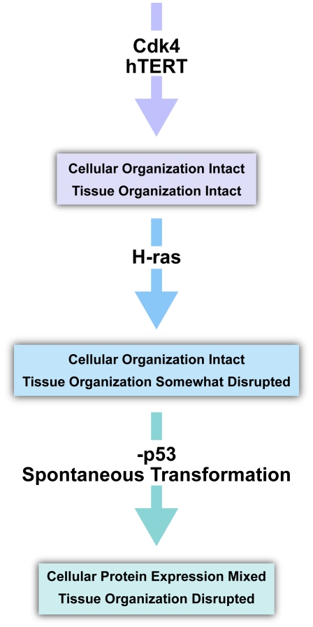
Background: Ras proteins affect both proliferation and expression of collagen-degrading enzymes, two important processes in cancer progression. Normal skin architecture is dependent both on the coordinated proliferation and stratification of keratinocytes, as well as the maintenance of a collagen-rich basement membrane. In the present studies we sought to determine whether expression of H-ras in skin keratinocytes would affect these parameters during the establishment and maintenance of an in vitro skin equivalent.

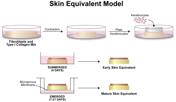
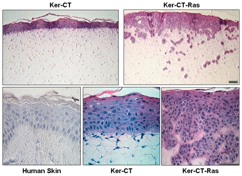
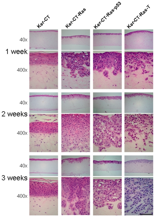
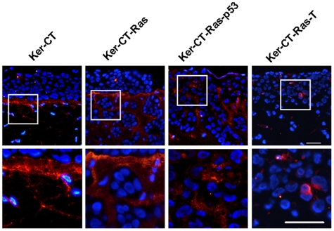
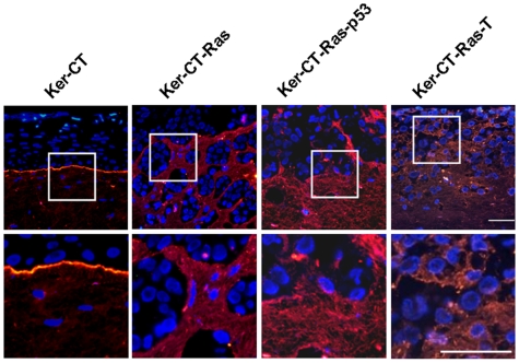
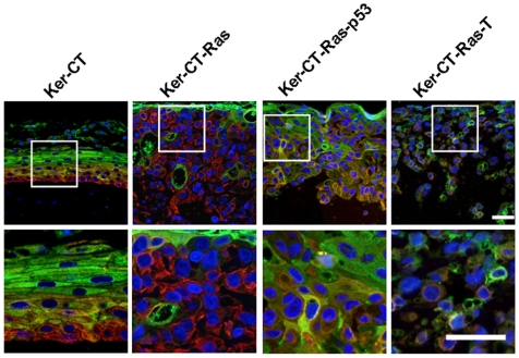
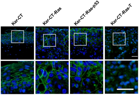
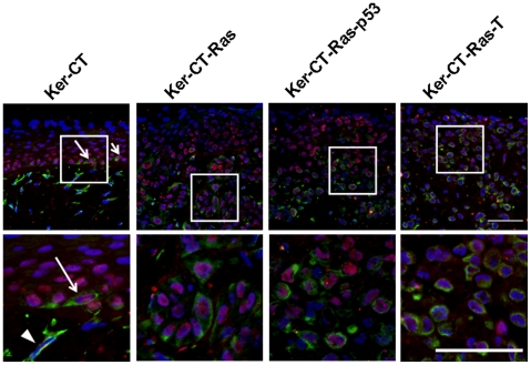
Methodology/principal findings: Previously described cdk4 and hTERT immortalized foreskin keratinocytes were engineered to express ectopically introduced H-ras. Skin equivalents, composed of normal fibroblast-contracted collagen gels overlaid with keratinocytes (immortal or immortal expressing H-ras), were prepared and incubated for 3 weeks. Harvested tissues were processed and sectioned for histology and antibody staining. Antigens specific to differentiation (involucrin, keratin-14, p63), basement-membrane formation (collagen IV, laminin-5), and epithelial to mesenchymal transition (EMT; e-cadherin, vimentin) were studied. Results showed that H-ras keratinocytes produced an invasive, disorganized epithelium most apparent in the lower strata while immortalized keratinocytes fully stratified without invasive properties. The superficial strata retained morphologically normal characteristics. Vimentin and p63 co-localization increased with H-ras overexpression, similar to basal wound-healing keratinocytes. In contrast, the cdk4 and hTERT immortalized keratinocytes differentiated similarly to normal unimmortalized keratinocytes.

Conclusions/significance: The use of isogenic derivatives of stable immortalized keratinocytes with specified genetic alterations may be helpful in developing more robust in vitro models of cancer progression.


Conflict of interest statement

Competing Interests: The authors have declared that no competing interests exist.

Figures

Figure 1
Figure 1. Diagram of the skin equivalent model.
Fibroblasts were mixed with collagen and allowed to contract over a period of 4–7 days. Keratinocytes were then plated, using a cloning ring, at a density of 200,000 cells/cm2. The cells were allowed 4 hours to settle and attach to the upper surface of the contracted collagen lattice. After 4 days of submerged culture, the skin equivalents were raised to the air/liquid interface through subsequent culturing in the upper chamber of a Transwell™ plate. Skin equivalents of emerged cultures were harvested at 7, 14, and 21 days.
Figure 2
Figure 2. Ker-CT-Ras keratinocytes produced a randomized, invasive epithelium.
While Ker-CT keratinocytes remain as a surface epithelium on the skin equivalent (upper left), Ker-CT-Ras keratinocytes appear to invade the dermal compartment (upper right). At higher magnification, normal human skin (lower left) and Ker-CT keratinocytes (lower center) produce histologically similar epithelium, while Ker-CT-Ras keratinocytes (lower right) lack the uniformity in the lower strata, although the stratum facing the air retained the ability to cornify. Scale bar: 30 µm.
Figure 3
Figure 3. Effect of time on epithelialization.
All skin equivalents demonstrated a stratified epithelium on the upper surface within one week. With H-Ras overexpression keratinocyte invasiveness typically occurred early and slowed with time. Similar results occurred with all H-Ras permutations. All but Ker-CT-Ras-T retained some ability to cornify. Upper images, 40× total magnification; lower images, 400× total magnification. Scale bar: 100 µm.
Figure 4
Figure 4. Collagen IV staining in 21-day skin equivalents.
Collagen IV staining (red) in Ker-CT skin equivalents increased during the 21 days of emerged culture. Non-linear collagen IV staining appeared in skin equivalents with H-Ras keratinocytes except for Ker-CT-Ras-T, which had little or no collagen IV staining. Lower images are insets of corresponding upper images. Scale bar: 40 µm.
Figure 5
Figure 5. Laminin-5 staining in 21-day skin equivalents.
Laminin-5 staining (red) in Ker-CT demonstrated a similar linear staining pattern as Collagen IV. In H-Ras keratinocytes, laminin-5 staining was more prevalent than collagen IV. Lower images are insets of corresponding upper images. Scale bar: 40 µm.
Figure 6
Figure 6. Double-staining of keratin-14 and involucrin in 21-day skin equivalents.
In Ker-CT keratinocytes keratin-14 (red) stained the lowermost layers, while involucrin (green) stained the uppermost layers. There was some evidence of co-localization (yellow to orange) in the intermediate layers. Upper staining of involucrin and lower staining of keratin-14 was partially retained in Ker-CT-Ras keratinocytes, but less so in Ker-CT-Ras-p53 keratinocytes. In Ker-CT-Ras-T keratinocytes there was little or no organization of the respective stains. In all tested cells, however, cellular co-localization of both proteins was rare. Lower images are insets of corresponding upper images. Scale bar: 40 µm.
Figure 7
Figure 7. E-cadherin staining in 21-day skin equivalents.
E-cadherin staining (green) in Ker-CT keratinocytes demonstrated the cellular connections reminiscent of stratum spinosum in human skin. Little staining was present on the basal surface of the basal layer. E-cadherin staining in H-Ras keratinocytes was less organized than Ker-CT although most of the interconnected cells were in the upper regions of the tissue. Ker-CT-Ras-T had little or no specific E-cadherin staining. Lower images are insets of corresponding upper images. Scale bar: 40 µm.
Figure 8
Figure 8. Double-staining of vimentin and p63 in 21-day skin equivalents.
p63 (red or pink) and vimentin (green) rarely co-localized in Ker-CT keratinocytes, except in occasional basal cells (arrow). Fibroblasts (arrowhead) were identified by the blue nucleus and green cytoplasm. More co-localization was seen in H-Ras keratinocytes, although primarily in the lower strata. Note the lack of either staining in superficial strata. Lower images are insets of corresponding upper images. Scale bar: 40 µm.
Figure 9
Figure 9. Combined analysis of protein overexpression effects on epithelial integrity.
CDK4 and hTERT expression in keratinocytes demonstrate little or no effect to overall tissue integrity or cellular protein staining profile. Upon expression of H-Ras the tissue integrity is affected but the cellular staining profile remains similar to cells without H-Ras expression. After p53 inhibition and transformation, both tissue integrity and cellular identity is lost. Tissue integrity loss may be due to the inappropriate environment itself.

Similar articles

Cited by

References

    1. Thiery JP, Sleeman JP. Complex networks orchestrate epithelial-mesenchymal transitions. Nat Rev Mol Cell Biol. 2006;7:131–142. - PubMed
    1. Guarino M. Epithelial-mesenchymal transition and tumor invasion. Int J Biochem Cell Biol. 2007;39:2153–2160. - PubMed
    1. Ridky TW, Khavari PA. Pathways sufficient to induce epidermal carcinogenesis. Cell Cycle. 2004;3:621–624. - PubMed
    1. Wennerberg K, Rossman KL, Der CJ. The Ras superfamily at a glance. J Cell Sci. 2005;118:843–846. - PubMed
    1. Shields JM, Pruitt K, McFall A, Shaub A, Der CJ. Understanding Ras: ‘it ain't over 'til it's over’. Trends Cell Biol. 2000;10:147–154. - PubMed

Publication types