. 2009 Nov 24;3(11):e553.
doi: 10.1371/journal.pntd.0000553.

Mycolactone gene expression is controlled by strong SigA-like promoters with utility in studies of Mycobacterium ulcerans and buruli ulcer

Affiliations

Mycolactone gene expression is controlled by strong SigA-like promoters with utility in studies of Mycobacterium ulcerans and buruli ulcer

Nicholas J Tobias et al. PLoS Negl Trop Dis. .

Erratum in

  • PLoS Negl Trop Dis. 2010;4(2). doi: 10.1371/annotation/1e664e6f-3fc8-4586-a84e-73c4ec4f783f

Abstract

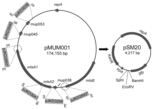
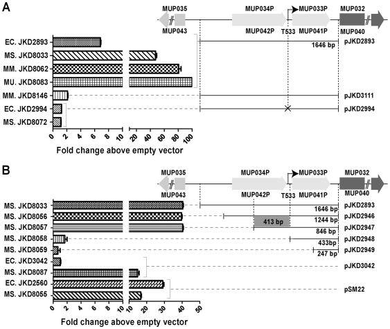
Mycolactone A/B is a lipophilic macrocyclic polyketide that is the primary virulence factor produced by Mycobacterium ulcerans, a human pathogen and the causative agent of Buruli ulcer. In M. ulcerans strain Agy99 the mycolactone polyketide synthase (PKS) locus spans a 120 kb region of a 174 kb megaplasmid. Here we have identified promoter regions of this PKS locus using GFP reporter assays, in silico analysis, primer extension, and site-directed mutagenesis. Transcription of the large PKS genes mlsA1 (51 kb), mlsA2 (7 kb) and mlsB (42 kb) is driven by a novel and powerful SigA-like promoter sequence situated 533 bp upstream of both the mlsA1 and mlsB initiation codons, which is also functional in Escherichia coli, Mycobacterium smegmatis and Mycobacterium marinum. Promoter regions were also identified upstream of the putative mycolactone accessory genes mup045 and mup053. We transformed M. ulcerans with a GFP-reporter plasmid under the control of the mls promoter to produce a highly green-fluorescent bacterium. The strain remained virulent, producing both GFP and mycolactone and causing ulcerative disease in mice. Mosquitoes have been proposed as a potential vector of M. ulcerans so we utilized M. ulcerans-GFP in microcosm feeding experiments with captured mosquito larvae. M. ulcerans-GFP accumulated within the mouth and midgut of the insect over four instars, whereas the closely related, non-mycolactone-producing species M. marinum harbouring the same GFP reporter system did not. This is the first report to identify M. ulcerans toxin gene promoters, and we have used our findings to develop M. ulcerans-GFP, a strain in which fluorescence and toxin gene expression are linked, thus providing a tool for studying Buruli ulcer pathogenesis and potential transmission to humans.


Conflict of interest statement

The authors have declared that no competing interests exist.

Figures

Figure 1
Figure 1. Schematic representation of the M. ulcerans Agy99 megaplasmid pMUM001, and the promoterless GFP vector pSM20.
The regions upstream of key genes involved in mycolactone biosynthesis cloned into pSM20 and the names of the resulting plasmid GFP-reporter constructs are indicated. The 8.4 kb duplicated region that spans the load modules of mlsA1 and mlsB and 1.6 kb upstream of each gene is circled. E = EcoRV, B = BamHI, S = SphI, ‘*’ indicates that this single construct represents the encircled duplicated regions upstream of mlsA1 and mlsB.
Figure 2
Figure 2. Summary of fluorescence data demonstrating the difference in promoter activity among strains containing GFP reporter constructs.
Shown are plasmid constructs with upstream regions from M. ulcerans mlsA1/mlsB (A) & (B). Also shown are the sigA promoter from M. bovis BCG (construct pJKD3042) (B) and the srp promoter from E. coli (construct pSM22) (B). Point mutations in putative promoter regions in the putative -10 motifs of promoter regions for mls sequences are marked by ‘X’. All values are expressed as fold changes above the strain containing the empty vector pSM20. Strains are identifiable by a two-letter prefix to the strain number EC - E. coli, MS - M. smegmatis, MM - M. marinum and MU - M. ulcerans. ‘*’ indicates this fluorescence is more than 100-fold higher than vector alone.
Figure 3
Figure 3. Histological study of serial mouse tail sections following mouse-tail infection with M. ulcerans-GFP.
(A) Fifty days after subcutaneous inoculation in the tail of 105 bacilli showing the beginning of ulceration. (B) Hematoxylin phloxine saffron staining on tissue section reveals massive necrosis. (C) Ziehl-Nieelsen staining showing many extracellular individual and clustered bacilli (arrowheads) associated with necrosis, and (D) bacilli expressing GFP. Scale bars: 0.8 cm (A), 100 µm (B, C, and D) and 40 µm (C and D insets).
Figure 4
Figure 4. Concentration of M. ulcerans-GFP within 4th instar mosquito larvae.
(A) Epifluorescence microscopy (x100 objective) demonstrating that the majority of total fluorescent bacteria (DAPI-staining, blue filter) are also GFP-expressing M. ulcerans (JKD8083) and M. marinum (JKD8062) (green filter). (B) Reduction of M. marinum and M. ulcerans within the water of the microcosms over the 4-week duration of the experiment as measured by qPCR. Results are the means and standard deviations of four biological repeats. (C) Bright field, red fluorescence, green fluorescence and composite images showing the accumulation of M. ulcerans around the mouth and mid-gut of a 4th instar Aedes camptorhynchus larva. Scale bar indicates 0.5 mm. Inset images are ×400 magnification. No specific green fluorescence was observed in larvae infected with M. marinum or uninfected controls. Red fluorescence images were included to show that green fluorescence was due to GFP expression and not autofluorescence.

Similar articles

Cited by

References

    1. Amofah G, Bonsu F, Tetteh C, Okrah J, Asamoa K, et al. Buruli ulcer in Ghana: results of a national case search. Emerg Infect Dis. 2002;8:167–170. - PMC - PubMed
    1. Debacker M, Aguiar J, Steunou C, Zinsou C, Meyers WM, et al. Mycobacterium ulcerans disease (Buruli ulcer) in rural hospital, Southern Benin, 1997–2001. Emerg Infect Dis. 2004;10:1391–1398. - PMC - PubMed
    1. Chauty A, Ardant MF, Adeye A, Euverte H, Guédénon A, et al. Promising clinical efficacy of streptomycin-rifampin combination for treatment of Buruli ulcer (Mycobacterium ulcerans disease). Antimicrob Agents Chemother. 2007;51:4029–4035. - PMC - PubMed
    1. van der Werf TS, van der Graaf WT, Tappero JW, Asiedu K. Mycobacterium ulcerans infection. Lancet. 1999;354:1013–1018. - PubMed
    1. Eddyani M, Ofori-Adjei D, Teugels G, De Weirdt D, Boakye D, et al. Potential role for fish in transmission of Mycobacterium ulcerans disease (Buruli ulcer): an environmental study. Appl Environ Microbiol. 2004;70:5679–5681. - PMC - PubMed

Publication types

MeSH terms