. 2009 Nov 11:15:2287-305.

Phenotypic variation and genotype-phenotype discordance in canine cone-rod dystrophy with an RPGRIP1 mutation

Affiliations

Phenotypic variation and genotype-phenotype discordance in canine cone-rod dystrophy with an RPGRIP1 mutation

Keiko Miyadera et al. Mol Vis. .

Abstract

Purpose: Previously, a 44 bp insertion in exon 2 of retinitis pigmentosa GTPase interacting protein 1 (RPGRIP1) was identified as the cause of cone-rod dystrophy 1 (cord1), a recessive form of progressive retinal atrophy (PRA) in the Miniature Longhaired Dachshund (MLHD), a dog model for Leber congenital amaurosis. The cord1 locus was mapped using MLHDs from an inbred colony with a homogeneous early onset disease phenotype. In this paper, the MLHD pet population was studied to investigate phenotypic variation and genotype-phenotype correlation. Further, the cord1 locus was fine-mapped using PRA cases from the MLHD pet population to narrow the critical region. Other dog breeds were also screened for the RGPRIP1 insertion.

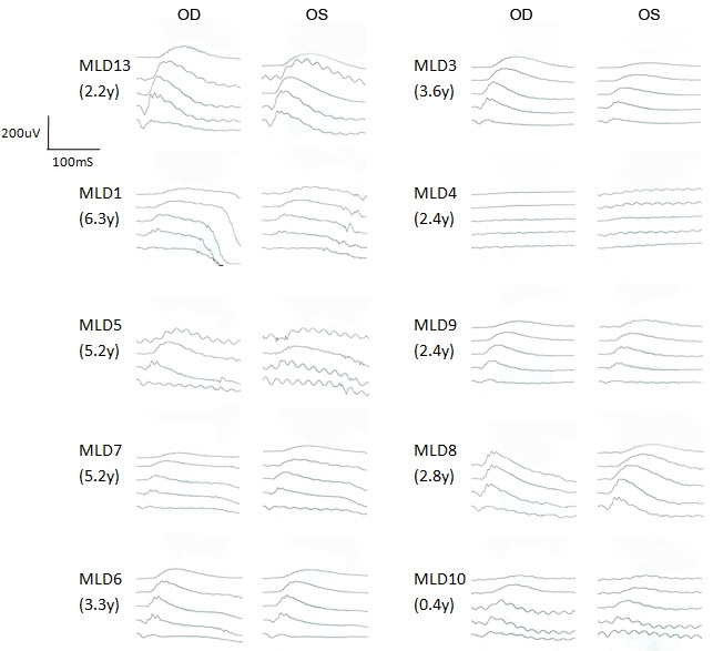
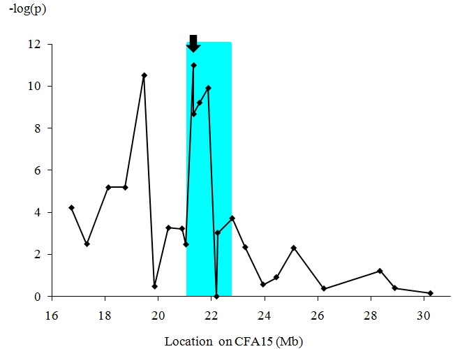
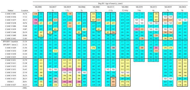
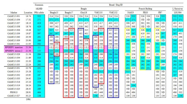
Methods: This study examined phenotypic variation in an MLHD pet population that included 59 sporadic PRA cases and 18 members of an extended family with shared environment and having six PRA cases. Ophthalmologic evaluations included behavioral abnormalities, responses to menace and light, fundoscopy, and electroretinography (ERG). The RPGRIP1 insertion was screened for in all cases and 200 apparently normal control MLHDs and in 510 dogs from 66 other breed. To fine-map the cord1 locus in the MLHD, 74 PRA cases and 86 controls aged 4 years or more were genotyped for 24 polymorphic markers within the previously mapped cord1 critical region of 14.15 Mb.

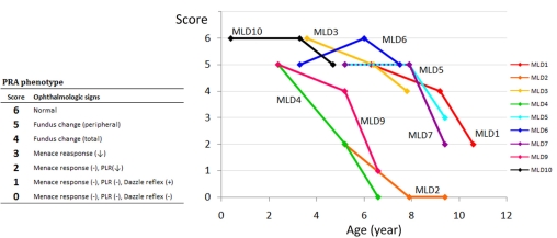
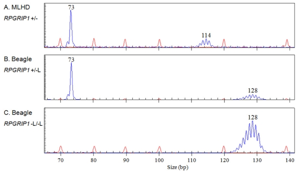
Results: Among sporadic PRA cases from the MLHD pet population, the age of onset varied from 4 months to 15 years old; MLHDs from the extended family also showed variable onset and rate of progression. Screening for the insertion in RPGRIP1 identified substantial genotype-phenotype discordance: 16% of controls were homozygous for the insertion (RPGRIP1(-/-)), while 20% of PRA cases were not homozygous for it. Four other breeds were identified to carry the insertion including English Springer Spaniels and Beagles with insertion homozygotes. The former breed included both controls and PRA cases, yet in the latter breed, cone ERG was undetectable in two dogs with no clinically apparent visual dysfunction. Notably, the insertion in the Beagles was a longer variant of that seen in the other breeds. Fine-mapping of the cord1 locus narrowed the critical region on CFA15 from 14.15 Mb to 1.74 Mb which still contains the RPGRIP1 gene.

Conclusions: Extensive phenotypic variations of onset age and progression rate were observed in PRA cases of the MLHD pet population. The insertion in RPGRIP1 showed the strongest association with the disease, yet additional as well as alternative factors may account for the substantial genotype-phenotype discordance.


Figures

Figure 1
Figure 1
Facial and fundoscopic characteristics of PRA. A: Typical PRA-affected cases show mydriasis and increased reflection from the fundus. B-D: MLHD fundus photographs of a control without visual dysfunction (B, 5y), moderately (C, 3.4y) and severely (D, 5y) affected PRA cases with blindness. Note the tapetal hyperreflectivity (C), the attenuation of the retinal vessels, the pale optic disk, and the pigmentation of the nontapetum (D).
Figure 2
Figure 2
Distribution of the age of PRA onset in sporadic MLHD cases. Presented are 48 sporadic PRA cases of MLHDs from the Japanese pet population. These are shown according to the age of onset. The dogs were already blind at the time of presentation and the age of onset was determined by the earliest possible sign of visual impairment noticed by the owner. An additional 11 sporadic PRA cases from Japan, used for mapping purposes, are not shown here since the information on the age of onset was uncertain or unknown. RPGRIP1 genotypes are denoted as follows: wildtype homozygote (+/+); heterozygote (+/−); insertion homozygote (−/−).
Figure 3
Figure 3
Pedigree of an extended family of MLHDs with shared environment. The family tree of Family K shows the proband (arrow), the dog’s three siblings from another pregnancy, and 14 other related members for which the retinal phenotype was examined over a period of 1.4 to 4.2 years. In the PRA affected cases, the age (y, years) under each symbol indicates the age of onset of behavioral (bold symbols) or fundoscopic (hatched symbols) abnormalities. In the dogs with no apparent visual dysfunction, the age corresponds to that at the last fundoscopic examination. Some members of the family were not examined in this study and are omitted from the pedigree. The following symbols are used: square, male; circle, female; bold, blind; hatched, abnormal fundus with no apparent visual dysfunction; white, no fundoscopic abnormality with no apparent visual dysfunction. RPGRIP1 genotypes are denoted as follows: wildtype homozygote, in blue (+/+); heterozygote, in yellow (+/−); insertion homozygote, in red (−/−).
Figure 4
Figure 4
Progression of the PRA phenotype in nine RPGRIP1−/− MLHDs an extended family with shared environment. The nine dogs shown are all RPGRIP1−/− with shared environment and were examined three times over a 4.2 year period. PRA phenotypes over a 4.2-year period are scored according to ophthalmologic abnormalities determined by fundoscopy, menace response, PLR, and dazzle reflex. A decreasing score indicates disease progression. Animals with scores below 3 are functionally blind and were included in the PRA affected group in the association analysis. MLD2, 5, and 7 and MLD4 and 9 are siblings.
Figure 5
Figure 5
Scotopic ERG in related MLHDs with no obvious sign of visual impairment. Scotopic ERG intensity series of 10 MLHDs from Family K at initial examination is shown. ERG recordings show scotopic responses with five increasing light intensity from top to bottom differing by 1 log cd/m2. The age in years (y) corresponds to the age when ERG was performed; at that time, none of the dogs showed apparent visual deficit. All dogs represented are RPGRIP1−/−, except MLD13 (RPGRIP1+/−). The data of the highest light intensity was not recorded in MLD5 or in the right eye (OD) of MLD8. OS indicates left eye.
Figure 6
Figure 6
PCR products containing the RPGRIP1 insertion were sized by capillary electrophoresis in the MLHD and the Beagle. Fragment sizing, by capillary electrophoresis of PCR products containing the RPGRIP1 insertion in a MLHD (A, RPGRIP1+/−) and two Beagles (B, RPGRIP1+/−L; C, RPGRIP1−L/−L). The 73 bp single blue peak corresponds to the wildtype allele (+), while the blue peaks centered on 114 bp and 128 bp represent the alleles with the insertion (- and -L); these two insertion alleles differ only in the number of adenines in the homopolymeric sequence. The red peaks correspond to the size standard.
Figure 7
Figure 7
ERG responses and fundus photograph of Beagles with the RPGRIP1 insertion variant. Bilateral scotopic (top) and photopic (middle) ERG recordings and fundus photograph of the left eye (OS; bottom) of three Beagles: a 5y RPGRIP1+/−L dog (A and D), a 2.8y RPGRIP−L/−L (B and E), and another 2.8y RPGRIP1−L/−L dog (C). Scotopic responses to a series of light stimuli are displayed with increasing light intensity from top to bottom differing by 1 log cd/m2 up to 18,400 cd/m2. The photopic response was recorded with 31 Hz flicker stimuli of 35,900 cd/m2. Note the apparently normal fundus appearance in the RPGRIP1−L/−L dog (E) with undetectable cone response (B). OD indicates right eye.
Figure 8
Figure 8
Fine mapping of the cord1 critical region. Twenty-four polymorphic markers on CFA15 were studied for the association with PRA in MLHDs. In total, 74 PRA cases and 86 controls were used for this fine mapping across the originally reported region of homozygosity [19]. Note that for the marker with the highest value at 21.34 Mb (RPGRIP1 insertion, arrow) the p-value is below the minimum detectable in the simulation, that is <1x10⁻¹¹. The aqua-highlighted area corresponds to the 1.74 Mb region of homozygosity shared by all PRA cases having two copies of the RPGRIP1 insertion.
Figure 9
Figure 9
Marker analysis in PRA-affected non-RPGRIP1−/− MLHDs. Haplotypes of the cord1 critical region on CFA15 were studied in 12 PRA-affected non RPGRIP1−/− MLHDs. The “common allele” refers to the combination of the most frequent allele observed in the RPGRIP1−/− PRA cases (47 sporadic cases and six cases from Family K). For each cell representing an allele in the dog studied, blue shading highlights genotypes identical to the haplotype associated with the cord1 insertion; yellow highlights alleles that are homozygous in the dog under study but differ from the common allele. The symbol (-) and (+) for the marker RPGRIP1 insertion each represents the RPGRIP1 insertion allele and the wildtype allele, respectively. ND indicates that the genotype was not determined.
Figure 10
Figure 10
Marker analysis in PRA affected RPGRIP1−/− MLHDs. Polymorphic markers in the cord1 critical region on CFA15 were analyzed in PRA affected RPGRIP1−/− MLHDs. The 12 dogs shown are representative of the haplotypes of 47 sporadic cases and six cases from Family K studied. Alleles shared by the majority of the dogs are highlighted in blue (referred to as “common allele” in Figure 9 and Figure 11) and other highlighted colors mark less common alleles. Variants in CAMC15.029 and 034 could be the result of mutation events, rather than recombination since the markers flanking them are the “common alleles.” Therefore, the region of homozygosity was delimited by markers CAMC15.041 and CAMC15.031. The symbol (-) for the marker RPGRIP1 insertion represents the RPGRIP1 insertion allele. ND indicates that the genotype was not determined.
Figure 11
Figure 11
Marker analysis in non-MLHD breeds with the RPGRIP1 insertion. Polymorphic markers in the cord1 critical region on CFA15 were analyzed in five RPGRIP1−L/−L Beagles, and in four other RPGRIP1+/− dogs (three French Bulldogs and a Labrador Retriever). The common allele denotes the combination of the most frequent allele observed in RPGRIP1−/− PRA cases of MLHDs (47 sporadic cases and six cases from Family K). Blue highlights mark genotypes identical to the common allele in the PRA-affected MLHDs. Haplotype blocks outlined in blue and red (Beagle) and yellow (French Bulldog) represent each breed-specific haplotype. Without taking into account the variation of the insertion length in RPGRIP1, the region of homozygosity shared between PRA-affected MLHDs and beagles both homozygous for an insertion in exon 2 of RPGRIP1 (RPGRIP1−/− and RPGRIP1−L/−L) was delimited by markers CAMC15.041 and CAMC15.029, which is highlighted in pink. The symbol (-) and (-L) for the marker RPGRIP1 insertion each represents the RPGRIP1 insertion allele and its variant while (+) represents the wildtype allele. ND indicates that the genotype was not determined.

Similar articles

Cited by

References

    1. Petersen-Jones S. Advances in the molecular understanding of canine retinal diseases. J Small Anim Pract. 2005;46:371–80. - PubMed
    1. Neitz J, Geist T, Jacobs GH. Color vision in the dog. Vis Neurosci. 1989;3:119–25. - PubMed
    1. Mowat FM, Petersen-Jones SM, Williamson H, Williams DL, Luthert PJ, Ali RR, Bainbridge JW. Topographical characterization of cone photoreceptors and the area centralis of the canine retina. Mol Vis. 2008;14:2518–27. - PMC - PubMed
    1. McGreevy P, Grassi TD, Harman AM. A strong correlation exists between the distribution of retinal ganglion cells and nose length in the dog. Brain Behav Evol. 2004;63:13–22. - PubMed
    1. Dekomien G, Runte M, Godde R, Epplen JT. Generalized progressive retinal atrophy of Sloughi dogs is due to an 8-bp insertion in exon 21 of the PDE6B gene. Cytogenet Cell Genet. 2000;90:261–7. - PubMed

Publication types

MeSH terms

LinkOut - more resources