

Matrix metalloproteinases in recurrent corneal melting associated with primary Sjörgen's syndrome
- PMID: 19936308
- PMCID: PMC2779063
Matrix metalloproteinases in recurrent corneal melting associated with primary Sjörgen's syndrome
Abstract
Purpose: To investigate the contribution of matrix metalloproteinases (MMPs) to recurrent corneal melting in keratoconjunctivitis sicca associated with primary Sjörgen's syndrome (pSS).
Methods: One native melted cornea and ten melted corneal grafts from two patients with severe pSS were used. The presence of MMPs (1, 2, 3, 7, 8, 9, and 13) was detected using indirect enzyme immunohistochemistry. The active forms of MMP 2 and 9 and MMP 3 and 7 were examined by gelatin and casein zymography, respectively. The concentrations of active MMP 1 were measured using an activity assay. Eleven unaffected corneas served as controls.
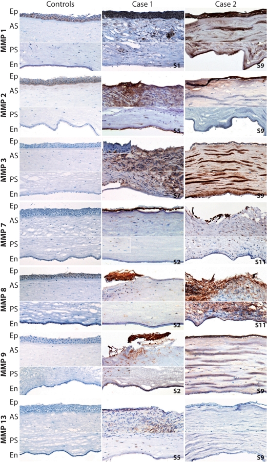
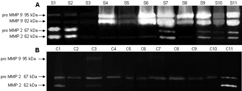
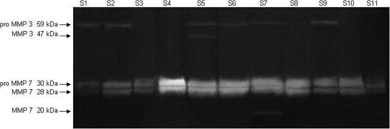
Results: The average values of the staining intensity revealed very intense MMP 1, intense MMP 2, 7, and 9 and moderate MMP 3 and 8 positivity, in the corneal epithelium of melted corneas. Intense MMP 1 and 9 staining, moderate MMP 2, 3, and 8 staining, and weak MMP 7 staining were found in the anterior stroma. The posterior stroma revealed intense MMP 1, moderate MMP 3 and 9, and weak MMP 2, 7, and 8 positivity. Immunostaining of the endothelium was moderate for MMP 9 and weak for MMP 1, 2, 3, 7, and 8. MMP 13 was negative in all but four melted specimens, where weak-to-moderate staining was found in the epithelium and stroma. Control corneas revealed moderate MMP 1 and 2 and weak MMP 8 staining in the epithelium, weak MMP 2 staining in the anterior stroma, and weak MMP 1 and 8 staining in the endothelium. Significantly elevated MMP 1 activity and extremely elevated MMP 9 activity were found in most of the tested pathological specimens, compared to healthy controls, where no activity of the two enzymes was present. Markedly elevated MMP 2 activity was found in 2 of 11 specimens, compared to normal tissue. The inactive form of MMP 3 was detected in half of the tested specimens, and inactive MMP 7 in all melted corneas. Active MMP 3 and 7 were found in one melted sample. Neither of these MMPs was found in any of the control specimens.
Conclusions: The increased expression and elevated activity of a wide range of MMPs in melted cornea samples from two patients diagnosed with pSS confirm that these enzymes contribute to the tissue destruction, leading to serious consequences such as corneal perforation and loss of vision.
Figures
Similar articles
-
Role of matrix metalloproteinases in recurrent corneal melting.Exp Eye Res. 2010 May;90(5):583-90. doi: 10.1016/j.exer.2010.02.002. Epub 2010 Feb 11. Exp Eye Res. 2010. PMID: 20153319
-
Expression of membrane-type matrix metalloproteinases 4, 5, and 6 in mouse corneas infected with P. aeruginosa.Invest Ophthalmol Vis Sci. 2001 Dec;42(13):3223-7. Invest Ophthalmol Vis Sci. 2001. PMID: 11726626
-
Overexpression of MMPs in Corneas Requiring Penetrating and Deep Anterior Lamellar Keratoplasty.Invest Ophthalmol Vis Sci. 2019 Apr 1;60(5):1734-1747. doi: 10.1167/iovs.18-25961. Invest Ophthalmol Vis Sci. 2019. PMID: 31022731 Free PMC article.
-
Is the corneal degradation in keratoconus caused by matrix-metalloproteinases?Clin Exp Ophthalmol. 2001 Dec;29(6):340-4. doi: 10.1046/j.1442-9071.2001.d01-17.x. Clin Exp Ophthalmol. 2001. PMID: 11778801 Review.
-
Proteinases of the cornea and preocular tear film.Vet Ophthalmol. 2007 Jul-Aug;10(4):199-206. doi: 10.1111/j.1463-5224.2007.00546.x. Vet Ophthalmol. 2007. PMID: 17565550 Review.
Cited by
-
Desiccating Stress-Induced MMP Production and Activity Worsens Wound Healing in Alkali-Burned Corneas.Invest Ophthalmol Vis Sci. 2015 Jul;56(8):4908-18. doi: 10.1167/iovs.15-16631. Invest Ophthalmol Vis Sci. 2015. PMID: 26225631 Free PMC article.
-
Corneal biomechanical alterations in patients with chronic ocular Graft Versus-Host Disease.PLoS One. 2019 Apr 25;14(4):e0213117. doi: 10.1371/journal.pone.0213117. eCollection 2019. PLoS One. 2019. PMID: 31022204 Free PMC article.
-
Time-dependent matrix metalloproteinases and tissue inhibitor of metalloproteinases expression change in fusarium solani keratitis.Int J Ophthalmol. 2016 Apr 18;9(4):512-8. doi: 10.18240/ijo.2016.04.06. eCollection 2016. Int J Ophthalmol. 2016. PMID: 27162721 Free PMC article.
-
MMP regulation of corneal keratocyte motility and mechanics in 3-D collagen matrices.Exp Eye Res. 2014 Apr;121:147-60. doi: 10.1016/j.exer.2014.02.002. Epub 2014 Feb 14. Exp Eye Res. 2014. PMID: 24530619 Free PMC article.
-
Alterations in corneal biomechanics underlie early stages of autoimmune-mediated dry eye disease.J Autoimmun. 2020 Nov;114:102500. doi: 10.1016/j.jaut.2020.102500. Epub 2020 Jun 18. J Autoimmun. 2020. PMID: 32565048 Free PMC article.
References
-
- Fox PC. Autoimmune diseases and Sjogren's syndrome: an autoimmune exocrinopathy. Ann N Y Acad Sci. 2007;1098:15–21. - PubMed
-
- Garcia-Carrasco M, Fuentes-Alexandro S, Escarcega RO, Salgado G, Riebeling C, Cervera R. Pathophysiology of Sjogren's syndrome. Arch Med Res. 2006;37:921–32. - PubMed
-
- Rehman HU. Sjogren's syndrome. Yonsei Med J. 2003;44:947–54. - PubMed
-
- Fox RI, Michelson P, Casiano CA, Hayashi J, Stern M. Sjogren's syndrome. Clin Dermatol. 2000;18:589–600. - PubMed
-
- Hansen A, Lipsky PE, Dorner T. Immunopathogenesis of primary Sjogren's syndrome: implications for disease management and therapy. Curr Opin Rheumatol. 2005;17:558–65. - PubMed
Publication types
MeSH terms
Substances
LinkOut - more resources
Full Text Sources
Medical