Short and long term axotomy-induced ERG changes in albino and pigmented rats
- PMID: 19936311
- PMCID: PMC2779069
Short and long term axotomy-induced ERG changes in albino and pigmented rats
Abstract
Purpose: To investigate the different components of full-field flash electroretinogram (ERG) responses in adult albino and pigmented rats at various time intervals following optic nerve transection (ONT).
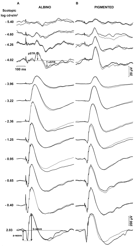
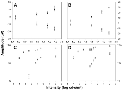
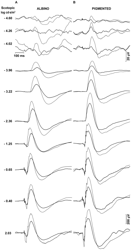
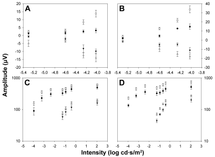
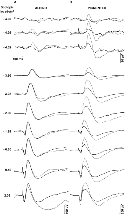
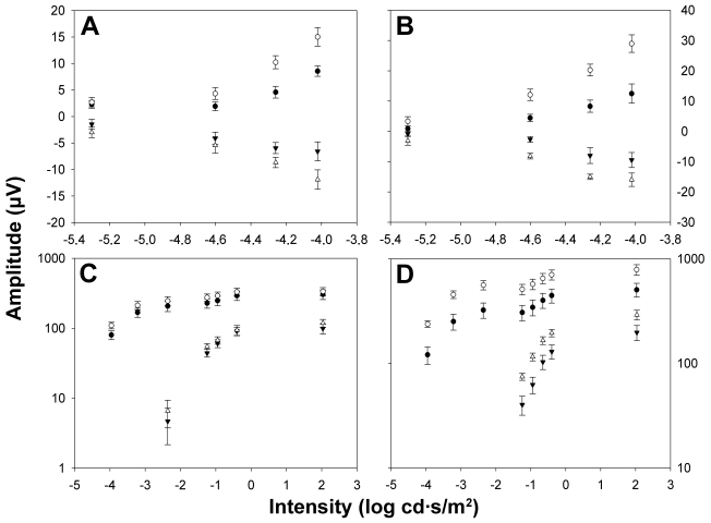
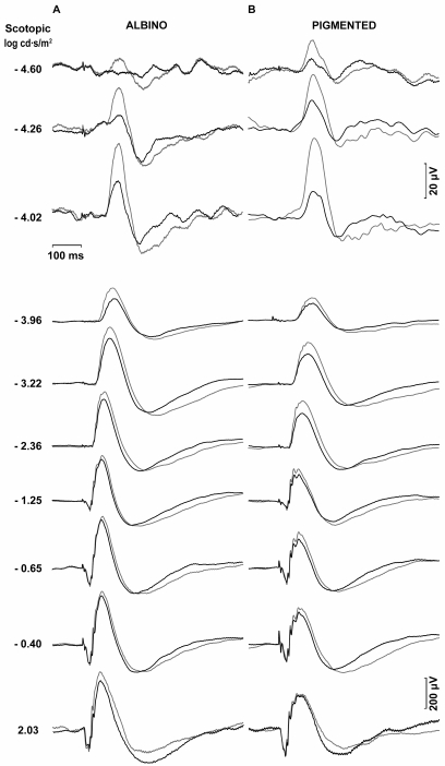
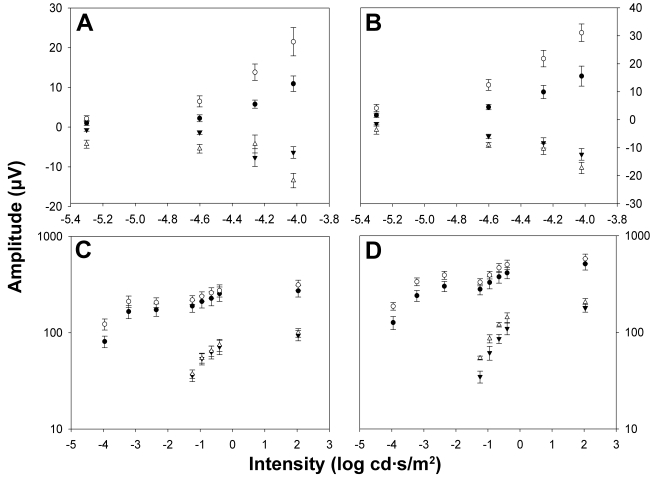
Methods: In adult Sprague-Dawley (SD, albino) and Piebald-Viral-Glaxo (PVG, pigmented) rats, the left optic nerve was transected intraorbitally to induce retinal ganglion cell (RGC) death. ERG responses were recorded simultaneously from both eyes beforehand and at 1, 2, 4, and 12 week intervals after ONT. The ERG a- and b-waves and the scotopic threshold responses (STR) were analyzed in scotopic conditions. White light stimuli of intensities ranging from 10(-6) to 10(-4) cd.s.m(-2) were used to record the positive and negative scotopic threshold responses (pSTR and nSTR), while stimulus light intensities ranging from 10(-4) to 10(2) cd.s.m(-2) were used to analyze the a- and b-wave amplitudes of standard ERG recordings.
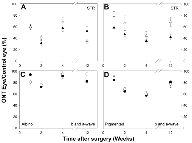
Results: In the albino rats, one week after intraorbital ONT, the STR mean amplitude decreased significantly, to approximately 60% of the values registered for the contralateral eye (p<0.05), which had not been operated on; standard ERG a- and b-waves showed a small reduction in amplitude-to approximately 85%. By two weeks after ONT, the STR mean amplitude was approximately 40% that of the contralateral eye, while the a- and b-wave amplitudes had further decreased to approximately 75%. Four weeks after ONT, the STR had fallen to 60% of that of the contralateral eyes, whereas the a- and b-waves reached values of approximately 90%. Twelve weeks after ONT, the STR remained significantly reduced to approximately 45%, whereas the a- and b-waves reached values of approximately 90%. In the pigmented rats, one week after intraorbital ONT, the mean amplitude had decreased significantly, to approximately 60% for the pSTR and to 80% for the nSTR of the values registered for the intact contralateral eye (p<0.05); while the standard ERG a- and b-waves showed a small reduction in amplitude to approximately 90%. Two weeks after ONT, the STR mean amplitude was approximately 55%, while the a- and b-wave amplitudes had further decreased to approximately 65%. Four weeks after ONT, the STR also was significantly reduced, to only 40%, whereas the a- and b-waves reached values of approximately 60%. Twelve weeks after ONT, the pSTR and nSTR remained significantly reduced to approximately 40% and 70%, respectively; whereas the a- and b-waves reached values of approximately 80%.
Conclusions: Optic nerve injury results in transient reductions of the major ERG components, the a- and b-waves, as well as permanent reductions of the early components of the ERG, the negative and positive scotopic threshold responses. Because ONT induces massive RGC loss, it is likely that permanent reduction in the STR represents a lack of the RGC population, thus highlighting the importance of the STR recordings as an electrophysiological tool for the assessment of RGC function.
Figures
Similar articles
-
ERG changes in albino and pigmented mice after optic nerve transection.Vision Res. 2010 Oct 12;50(21):2176-87. doi: 10.1016/j.visres.2010.08.014. Epub 2010 Aug 19. Vision Res. 2010. PMID: 20727908
-
Ganglion cell contributions to the rat full-field electroretinogram.J Physiol. 2004 Feb 15;555(Pt 1):153-73. doi: 10.1113/jphysiol.2003.052738. Epub 2003 Oct 24. J Physiol. 2004. PMID: 14578484 Free PMC article.
-
7,8-Dihydroxiflavone Maintains Retinal Functionality and Protects Various Types of RGCs in Adult Rats with Optic Nerve Transection.Int J Mol Sci. 2021 Oct 30;22(21):11815. doi: 10.3390/ijms222111815. Int J Mol Sci. 2021. PMID: 34769247 Free PMC article.
-
[Nonarteritic ischemic optic neuropathy animal model and its treatment applications].Nippon Ganka Gakkai Zasshi. 2014 Apr;118(4):331-61. Nippon Ganka Gakkai Zasshi. 2014. PMID: 24864434 Review. Japanese.
-
The Electroretinogram: ERG.2001 May 1 [updated 2007 Jun 27]. In: Kolb H, Fernandez E, Jones B, Nelson R, editors. Webvision: The Organization of the Retina and Visual System [Internet]. Salt Lake City (UT): University of Utah Health Sciences Center; 1995–. 2001 May 1 [updated 2007 Jun 27]. In: Kolb H, Fernandez E, Jones B, Nelson R, editors. Webvision: The Organization of the Retina and Visual System [Internet]. Salt Lake City (UT): University of Utah Health Sciences Center; 1995–. PMID: 21413407 Free Books & Documents. Review.
Cited by
-
Retinal compensatory changes after light damage in albino mice.Mol Vis. 2012;18:675-93. Epub 2012 Mar 24. Mol Vis. 2012. PMID: 22509098 Free PMC article.
-
A Chronic Ocular-Hypertensive Rat Model induced by Injection of the Sclerosant Agent Polidocanol in the Aqueous Humor Outflow Pathway.Int J Mol Sci. 2019 Jun 29;20(13):3209. doi: 10.3390/ijms20133209. Int J Mol Sci. 2019. PMID: 31261943 Free PMC article.
-
Activation of adenosine A3 receptor protects retinal ganglion cells from degeneration induced by ocular hypertension.Cell Death Dis. 2020 May 27;11(5):401. doi: 10.1038/s41419-020-2593-y. Cell Death Dis. 2020. PMID: 32461578 Free PMC article.
-
The retina of the lab rat: focus on retinal ganglion cells and photoreceptors.Front Neuroanat. 2022 Sep 23;16:994890. doi: 10.3389/fnana.2022.994890. eCollection 2022. Front Neuroanat. 2022. PMID: 36213609 Free PMC article. Review.
-
Using the Electroretinogram to Assess Function in the Rodent Retina and the Protective Effects of Remote Limb Ischemic Preconditioning.J Vis Exp. 2015 Jun 9;(100):e52658. doi: 10.3791/52658. J Vis Exp. 2015. PMID: 26131649 Free PMC article.
References
-
- Dowling JE. The Retina. An Approachable part of the brain. Cambridge, MA: The Belknap Press of Harvard University Press; 1987.
-
- Frishman LJ. Origins of the Electroretinogram. In: Heckenlively JR, Arden GB, editors. Principles and Practice of Clinical Electrophysiology of Vision. 2nd ed. Cambridge, MA: Massachusetts Institute of Technology (MIT); 2006. p. 139–84.
-
- Wachtmeister L. Oscillatory potentials in the retina: what do they reveal. Prog Retin Eye Res. 1998;17:485–521. - PubMed
-
- Sieving PA, Frishman LJ, Steinberg RH. Scotopic threshold response of proximal retina in cat. J Neurophysiol. 1986;56:1049–61. - PubMed
-
- Frishman LJ, Steinberg RH. Intraretinal analysis of the threshold dark-adapted ERG of cat retina. J Neurophysiol. 1989;61:1221–32. - PubMed
Publication types
MeSH terms
LinkOut - more resources
Full Text Sources
Other Literature Sources