. 2010 Feb;67(4):641-53.
doi: 10.1007/s00018-009-0201-5. Epub 2009 Nov 21.

Reelin is a platelet protein and functions as a positive regulator of platelet spreading on fibrinogen

Affiliations

Reelin is a platelet protein and functions as a positive regulator of platelet spreading on fibrinogen

Wei-Lien Tseng et al. Cell Mol Life Sci. 2010 Feb.

Abstract

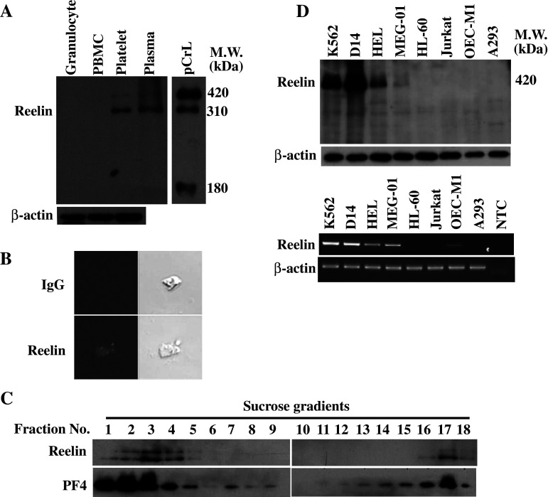
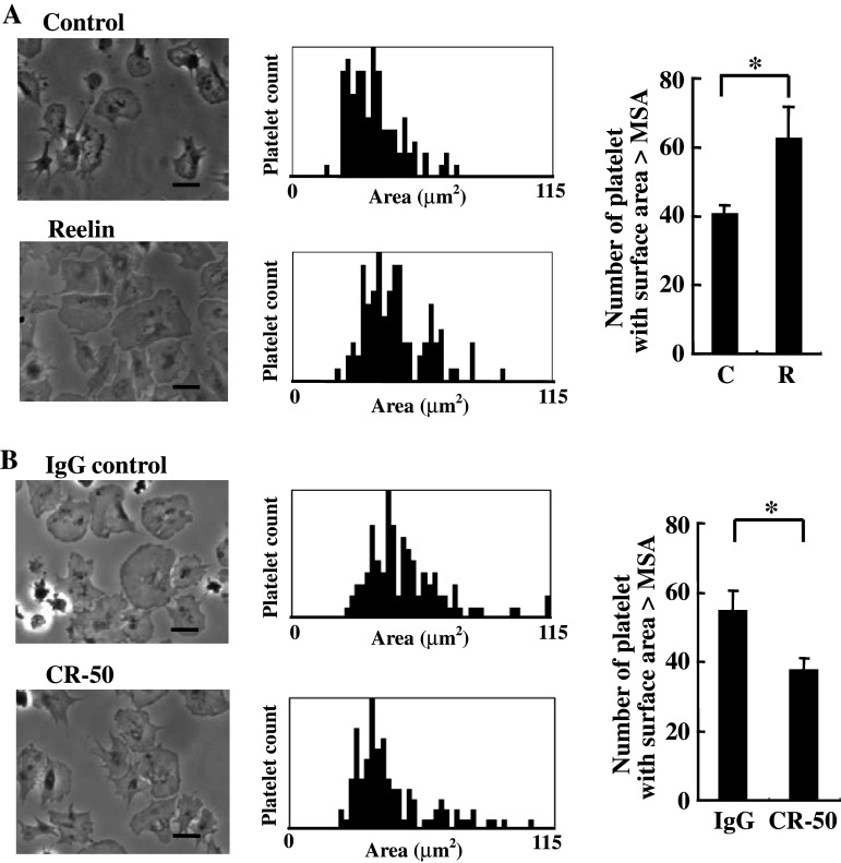
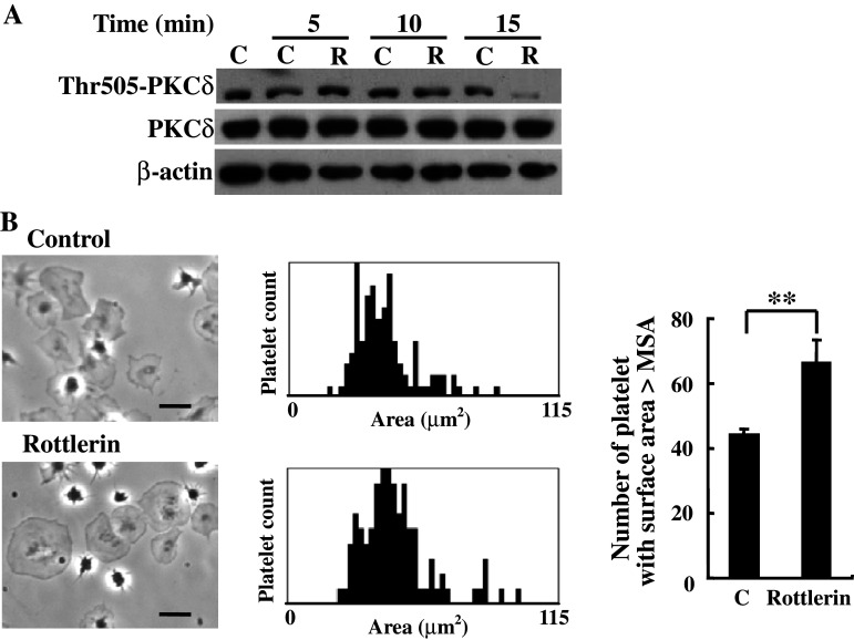
Abnormalities of platelet functions have been linked to reelin-impaired neuronal disorders. However, little attention has been given to understanding the interplay between reelin and platelet. In this study, reelin was found to present in the human platelets and megakaryocyte-like leukemic cells. Reelin-binding assays revealed that extracellular reelin can interact with platelets through the receptor belonging to the low density lipoprotein receptor gene family. The reelin-to-platelet interactions enhance platelet spreading on fibrinogen concomitant with the augmentation of lamellipodia formation and F-actin bundling. In contrast, reelin has no effect on integrin alphaIIbbeta3 activation and agonist-induced platelet aggregation. Molecular analysis revealed that the up-regulation of Rac1 activity and the inhibition of protein kinase C delta-Thr505 phosphorylation are important for reelin-mediated enhancement of platelet spreading on fibrinogen. These findings demonstrate for the first time that reelin is present in platelets and the reelin-to-platelet interactions play a novel role in platelet signaling and functions.


Figures

Fig. 1
Fig. 1
Reelin expression in the peripheral blood cells and various leukemic and cancer cells. a Expression of reelin in peripheral blood cells. The total lysates of granulocyte, PBMC, platelet and plasma were subjected to Western blot analysis with mAb142. The total lysate of 293T cells transfected with reelin expression plasmid pCrl was also included for comparison. b Immunofluorescence staining of reelin in platelets. Immunofluorescence staining with mAb142 or the IgG control was performed with platelets cytospun on a glass slide. The fluorescence signal was observed by confocal microscopy. c Subcellular localization of reelin in human platelets. Platelet homogenates (2 × 109) were separated by sucrose-density-gradient (30–60%) centrifugation. Eighteen fractions (700 μl each) were collected from the top, and aliquots were subjected to Western blot analysis using anti-reelin (mAb142) and anti-PF4 antibodies, respectively. d Expression of reelin in various leukemic and cancer cell lines. Total cell lysates and total RNA from the indicated cell lines were subjected to Western blot analysis using mAb142 (upper panel) and RT-PCR using reelin-specific primers (lower panel), respectively. The expression of β-actin was used as a control for equal protein loading and quality control of total RNA isolation. NTC, no template control
Fig. 2
Fig. 2
Human platelets interact with soluble and immobilized reelin. a The interaction between platelet and immobilized reelin. The washed platelets (500 μl) at a density of 3 × 108/ml were added to the coverslips pre-coated with purified reelin or the corresponding control for 30 min at 37°C. After washing and fixation, the binding of platelets on immobilized reelin was observed by phase contrast microscopy (upper panel). Note the filopodia-like protrusion for the platelets in the enlarged image. The relative number of platelet adhesions and the percentage of adhered platelets with filopodia-like protrusions were determined (lower panel). The data represent the mean ± SD of three independent experiments. **P < 0.01 and *P < 0.05 when compared with the control-treated coverslip. Length of black bar = 10 μm. C control, R reelin. b GST-RAP attenuates platelet binding on immobilized reelin. Solid phase binding assays were performed as described in the presence of GST or GST-RAP (40 μg/ml). The platelets were observed by phase contrast microscopy, and the relative number of platelets adhered on the control or reelin-coated coverslips was determined. The data represent the mean ± SD of three independent experiments. **P < 0.01 when compared with the platelet binding on immobilized reelin in the presence of GST protein. Length of black bar = 10 μm. c The 420-kDa reelin is involved in reelin-to-platelet interaction. The washed platelets (3 × 108/ml) were incubated with the purified reelin or the corresponding control for 2 h at 4°C. After several washes with 1× PBS, the platelet lysates were subjected to Western blot analysis with mAb142. The protein bands corresponding to the endogenous (endo) and exogenous (exo) reelin were shown. The expression of β-actin was used as a control for equal protein loading
Fig. 3
Fig. 3
Effects of reelin on platelet aggregation and integrin αIIbβ3 activation. a Reelin does not induce platelet aggregation. The washed platelets (left panel) and PRP (right panel) were subjected to platelet aggregation analysis in the presence of purified recombinant reelin or the corresponding control. The platelet aggregation curves were obtained by using a platelet aggregometer connected to the PowerLab data acquisition and recording system. b Reelin does not induce integrin αIIbβ3 activation. The platelets with the indicated treatments were incubated with the anti-PAC-1 (20 μl) antibody and subjected to flow cytometry analysis. A total of 10,000 events were determined. Similar results were obtained in three independent experiments. c Reelin does not modulate agonist-induced platelet aggregation. The washed platelets were incubated with purified recombinant reelin or the corresponding control for 1 min, and platelet aggregation was induced by the indicated concentrations of agonists. The platelet aggregation curves were recorded as described in a
Fig. 4
Fig. 4
Reelin enhances platelet spreading on fibrinogen. (a and b) The washed platelets (3 × 108/ml) were incubated with the purified reeelin or the corresponding control for 15 min at 37°C (a). Alternatively, the washed platelets were incubated with the purified reelin in the presence of reelin function blocking antibody CR-50 (20 μg/ml) or IgG control antibody for 15 min at 37°C (b). The assays of platelet spreading on fibrinogen were then performed and quantified as described in the “Materials and methods.” Platelet spreading was observed by phase contrast microscopy (left panel). The surface area for a total of 100 platelets in a representative experiment was plotted (middle panel), and the number of platelets with a surface area larger than MSA for the platelets in the control (a) or the IgG control (b) were determined (right panel). The data represent the mean ± SD of three independent experiments. *P < 0.05 when compared with the control (a) or the IgG-control (b). Length of black bar = 10 μm
Fig. 5
Fig. 5
Reelin moderates F-actin distribution and up-regulates platelet Rac1 activity. a Reelin does not affect the levels of F-actin for resting and thrombin-stimulating platelets. The washed platelets were pre-treated with purified recombinant reelin or the corresponding control for 30 min. The platelets were either untreated (resting) or were stimulated with thrombin (1 U/ml) for 1 min. F-actin staining with FITC-conjugated phalloidin was then performed and was quantified by flow cytometry. A total of 10,000 events were determined. Similar results were obtained in three independent experiments. b Reelin modifies F-actin distribution. The washed platelets were treated with purified recombinant reelin or the corresponding control for 15 min. The platelets were then spread on a fibrinogen-coated coverslip for 30 min. The distribution of F-actin was observed by staining platelets with FITC-conjugated phalloidin and observed by phase contrast microscopy. c Reelin up-regulates platelet Rac1 activity. The washed platelets were treated with purified recombinant reelin or the corresponding control for the indicated time. Analysis of Rac1 activity was then performed, and the amount of active Rac1 (GTP-Rac1) was analyzed by Western blot with the anti-Rac1 antibody. At the same time, Rac1 expression in the total platelet lysates was analyzed for loading control. Three independent experiments were performed with essentially similar results. d NSC23766 reverses reelin-mediated platelet spreading on fibrinogen. The washed platelets were incubated with purified recombinant reelin in the presence or absence of the Rac1 inhibitor NSC23766 (50 μM). Platelet spreading on fibrinogen was then performed as described in the “Materials and methods.” The patterns of platelet spreading were observed by phase contrast microscopy (left panel), and the surface area for a total of 100 platelets in a representative experiment was plotted (middle panel). The number of platelets with a surface area larger than MSA of the reelin-treated group was determined (right panel). The data represent the mean ± SD of three independent experiments. **P < 0.01 when compared with the reelin-treated group. C control, R reelin, N NSC23766. Length of black bar = 10 μm
Fig. 6
Fig. 6
Involvement of PKCδ in reelin signaling and platelet spreading on fibrinogen. a Reelin inhibits PKCδ phosphorylation at Thr505. The washed platelets were treated with recombinant reelin or the corresponding control for the indicated time. The platelet lysates were subjected to Western blot analysis with the anti-phospho-Thr505-PKCδ, anti-PKCδ and anti-β-actin antibodies, respectively. b Inhibition of PKCδ enhances platelet spreading on fibrinogen. Platelet spreading on fibrinogen was performed in the presence or absence of PKCδ inhibitor rottlerin (5 μM). The patterns of platelet spreading were observed by phase contrast microscopy (left panel) and the surface area for a total of 100 platelets in a representative experiment was plotted (middle panel). The number of platelets with a surface area larger than MSA of the control was determined (right panel). The data represent the mean ± SD of three independent experiments. **P < 0.01 when compared with the control-treated group. Length of black bar = 10 μm

References

    1. D’Arcangelo G, Miao GG, Chen SC, Soares HD, Morgan JI, Curran T. A protein related to extracellular matrix proteins deleted in the mouse mutant reeler. Nature. 1995;374:719–723. doi: 10.1038/374719a0. - DOI - PubMed
    1. Quattrocchi CC, Wannenes F, Persico AM, Ciafre SA, D’Arcangelo G, Farace MG, Keller F. Reelin is a serine protease of the extracellular matrix. J Biol Chem. 2002;277:303–309. doi: 10.1074/jbc.M106996200. - DOI - PubMed
    1. Assadi AH, Zhang G, Beffert U, McNeil RS, Renfro AL, Niu S, Quattrocchi CC, Antalffy BA, Sheldon M, Armstrong DD, Wynshaw-Boris A, Herz J, D’Arcangelo G, Clark GD. Interaction of reelin signaling and Lis1 in brain development. Nat Genet. 2003;35:270–276. doi: 10.1038/ng1257. - DOI - PubMed
    1. Hiesberger T, Trommsdorff M, Howell BW, Goffinet A, Mumby MC, Cooper JA, Herz J. Direct binding of Reelin to VLDL receptor and ApoE receptor 2 induces tyrosine phosphorylation of disabled-1 and modulates tau phosphorylation. Neuron. 1999;24:481–489. doi: 10.1016/S0896-6273(00)80861-2. - DOI - PubMed
    1. Bock HH, Herz J. Reelin activates SRC family tyrosine kinases in neurons. Curr Biol. 2003;13:18–26. doi: 10.1016/S0960-9822(02)01403-3. - DOI - PubMed

Publication types

MeSH terms