

Activating mutations of the GNAQ gene: a frequent event in primary melanocytic neoplasms of the central nervous system
- PMID: 19936769
- PMCID: PMC2831181
- DOI: 10.1007/s00401-009-0611-3
Activating mutations of the GNAQ gene: a frequent event in primary melanocytic neoplasms of the central nervous system
Abstract
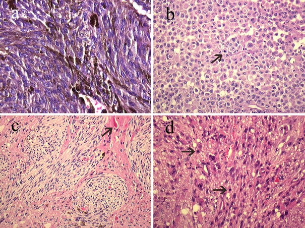
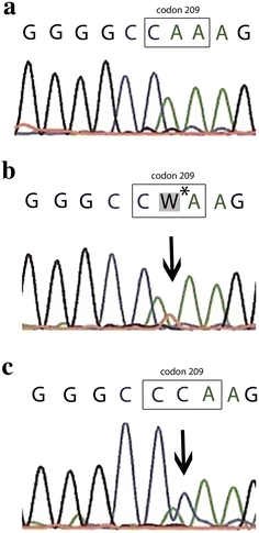
Primary melanocytic neoplasms of the central nervous system (CNS) are uncommon neoplasms derived from melanocytes that normally can be found in the leptomeninges. They cover a spectrum of malignancy grades ranging from low-grade melanocytomas to lesions of intermediate malignancy and overtly malignant melanomas. Characteristic genetic alterations in this group of neoplasms have not yet been identified. Using direct sequencing, we investigated 19 primary melanocytic lesions of the CNS (12 melanocytomas, 3 intermediate-grade melanocytomas, and 4 melanomas) for hotspot oncogenic mutations commonly found in melanocytic tumors of the skin (BRAF, NRAS, and HRAS genes) and uvea (GNAQ gene). Somatic mutations in the GNAQ gene at codon 209, resulting in constitutive activation of GNAQ, were detected in 7/19 (37%) tumors, including 6/12 melanocytomas, 0/3 intermediate-grade melanocytomas, and 1/4 melanomas. These GNAQ-mutated tumors were predominantly located around the spinal cord (6/7). One melanoma carried a BRAF point mutation that is frequently found in cutaneous melanomas (c.1799 T>A, p.V600E), raising the question whether this is a metastatic rather than a primary tumor. No HRAS or NRAS mutations were detected. We conclude that somatic mutations in the GNAQ gene at codon 209 are a frequent event in primary melanocytic neoplasms of the CNS. This finding provides new insight in the pathogenesis of these lesions and suggests that GNAQ-dependent mitogen-activated kinase signaling is a promising therapeutic target in these tumors. The prognostic and predictive value of GNAQ mutations in primary melanocytic lesions of the CNS needs to be determined in future studies.
Figures
References
-
- Balmaceda CM, Fetell MR, O’Brien JL, Housepian EH. Nevus of Ota and leptomeningeal melanocytic lesions. Neurology. 1993;43:381–386. - PubMed
-
- Bisceglia M, Carosi I, Fania M, Di CA, Lomuto M. Nevus of Ota. Presentation of a case associated with a cellular blue nevus with suspected malignant degeneration and review of the literature. Pathologica. 1997;89:168–174. - PubMed
-
- Brat DJ, Perry A. Melanocytic lesions. In: Louis DN, Ohgaki H, Wiestler OD, Cavenee WK, editors. WHO classification of tumours of the central nervous system. 4. Lyon: IARC; 2007. pp. 181–183.
MeSH terms
Substances
LinkOut - more resources
Full Text Sources
Other Literature Sources
Medical
Research Materials
Miscellaneous
