Review
. 2010 Jan;200(3-4):197-222.
doi: 10.1007/s00221-009-2054-4.

Internal models and neural computation in the vestibular system

Affiliations
Review

Internal models and neural computation in the vestibular system

Andrea M Green et al. Exp Brain Res. 2010 Jan.

Abstract

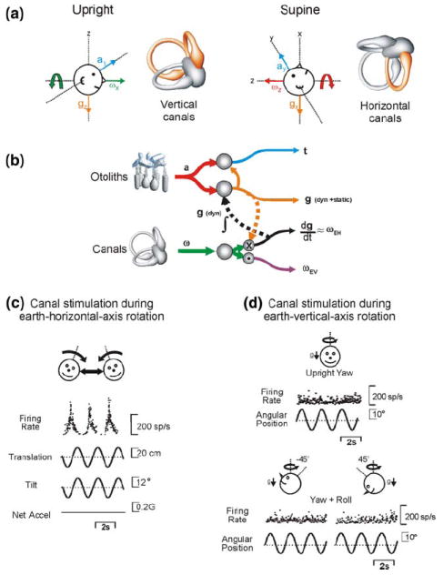
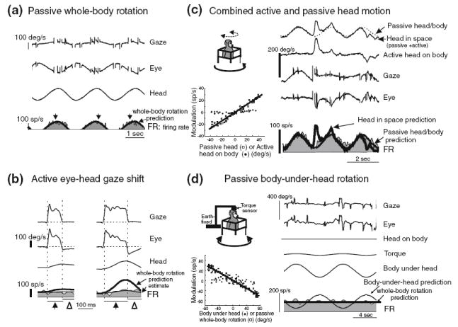
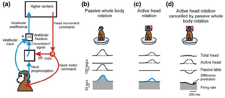
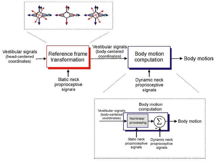
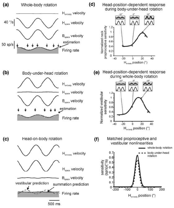
The vestibular system is vital for motor control and spatial self-motion perception. Afferents from the otolith organs and the semicircular canals converge with optokinetic, somatosensory and motor-related signals in the vestibular nuclei, which are reciprocally interconnected with the vestibulocerebellar cortex and deep cerebellar nuclei. Here, we review the properties of the many cell types in the vestibular nuclei, as well as some fundamental computations implemented within this brainstem-cerebellar circuitry. These include the sensorimotor transformations for reflex generation, the neural computations for inertial motion estimation, the distinction between active and passive head movements, as well as the integration of vestibular and proprioceptive information for body motion estimation. A common theme in the solution to such computational problems is the concept of internal models and their neural implementation. Recent studies have shed new insights into important organizational principles that closely resemble those proposed for other sensorimotor systems, where their neural basis has often been more difficult to identify. As such, the vestibular system provides an excellent model to explore common neural processing strategies relevant both for reflexive and for goal-directed, voluntary movement as well as perception.


Figures

Fig. 1
Fig. 1
The sensorimotor processing underlying eye movement generation in the RVOR. a Angular velocity signals from the semicircular canals are processed by an inverse dynamic model of the eye plant before being conveyed onto extraocular motoneurons (MNs). b Predicted frequency response characteristics of the RVOR either with (red curve) or without (blue curve) processing by the inverse model in a. Actual RVOR responses extend to lower frequencies than predicted by the blue curve but closely follow the red curve, demonstrating that sensory signals are processed by an inverse eye plant model. c Parallel-pathway implementation of the inverse dynamic model originally proposed by Skavenski and Robinson (1973). The inverse model (gray shaded box) is constructed by summing a weighted combination of angular velocity (top) and integrated angular velocity signals (bottom). An internal estimate of desired eye position (E*) is presumed to exist at the output of the neural integrator (∫). d Distributed feedback implementation of the inverse model proposed by Galiana and colleagues (Galiana and Outerbridge 1984; Galiana 1991). The required neural integration was proposed to be implemented via positive feedback loops through a forward model of the eye plant. Modified and reprinted with permission from Green et al. 2007
Fig. 2
Fig. 2
Premotor eye-movement-sensitive cell types implicated in the brainstem RVOR pathways. a Burst-tonic (PH–BT) neuron recorded in the PH. Modified and reprinted with permission from McFarland and Fuchs (1992). b Eye-contralateral (Type I) position-vestibular-pause (PVP) neuron recorded in the rostral medial VN. c Eye-contralateral eye-head (EH) neuron recorded in the rostral medial VN. Modified and reprinted with permission from Scudder and Fuchs (1992)
Fig. 3
Fig. 3
Schematic illustration of two hypotheses for the dynamic processing in the TVOR. a Distributed dynamic processing hypothesis, whereby the internal model is not fully implemented in the TVOR pathways. Otolith signals are processed by only the neural integrator portion (dashed box) of the inverse model that converts linear acceleration signals into velocity. In this scheme, the dynamic characteristics of the eye plant remain uncompensated and the plant dynamics contribute to shaping the reflex at higher frequencies, where the TVOR exhibits a robust response. b Common internal model hypothesis. Otolith signals, encoding linear acceleration, are presumed to be “prefiltered” before converging onto a common inverse model used to convert desired eye velocity signals into appropriate motor commands. Modified and reprinted with permission from Green et al (2007)
Fig. 4
Fig. 4
Evidence that tonic and burst-tonic cells in the PH and adjacent medial VN (PH–BT cells) represent the output of a common inverse model. a Comparison of the dynamic characteristics of PH–BT cells with those of extraocular motoneurons. Neural response gain (left) and phase (right) relative to eye position of PH–BT cells (triangles, solid lines) and motoneurons in the abducens (circles, dashed lines) and oculomotor (squares, dotted lines) nuclei are plotted during rotation (red curves) and translation (blue curves) as a function of frequency. Phases are expressed relative to the preferred eye movement direction of the cell. b Schematic illustration of the dynamic processing for eye movement generation in which PH–BT cells represent the output of the inverse dynamic model. Although the PH–BT cell population is illustrated outside the box labeled “inverse model” for simplicity, these cells can also be considered to contribute as an integral part of the model through feedback interconnections (dotted line) with other premotor neural populations including PVP and EH neurons. All three cell types project to the motoneuron population, MN. PH–BT cells are postulated to distribute an estimate of the motor command signal to other brain areas (e.g., the cerebellar flocculus) that potentially implement a forward model of the eye plant (or of the gaze system in general). The estimated motor response at the output of such a forward model can be compared with the desired eye movement to help refine the motor command signal. Modified and replotted with permission from Green et al. 2007
Fig. 5
Fig. 5
Evidence for a neural resolution to the tilt/translation ambiguity. Responses of an otolith afferent (a) and a mainly translation-coding rostral VN neuron (b) recorded during four tilt/translation stimulus combinations in darkness. Modified and replotted with permission from Angelaki et al. 2004. c Summary of how well brainstem and cerebellar neurons discriminate tilts and translations. The plot illustrates Z-transformed partial correlation coefficients for the fits of individual cell responses with a translation-coding model and a net acceleration (afferent-like) model. NU Purkinje cells (blue filled circles), rFN cells (orange up triangles), VN neurons (green down triangles) are compared with primary otolith afferents (red filled squares). Dashed lines divide the plots into an upper-left region where cell responses were significantly better fit (p < 0.01) by the translation-coding model and a lower-right region where neurons were significantly better fit by the net acceleration model. The intermediate area represents a region where cells were not significantly better fit by either model. Notice that unlike the distributed representation of VN and rFN cells, most NU cells are best fit by a translation-coding model (i.e., blue circles in upper left region). Modified and replotted with permission from Yakusheva et al. (2007) and Angelaki and Cullen (2008)
Fig. 6
Fig. 6
A reference frame transformation of canal signals is required to discriminate tilts and translations in 3D. a Only rotations about an earth-horizontal axis (but not an earth-vertical axis) reorient the head relative to gravity (e.g., thick green and red arrows). As a result, resolution of the tilt/translation ambiguity relies on different combinations of canal signals depending on one’s current head orientation. For example, when upright (left), roll rotation about the head x-axis (green arrow) stimulates the otoliths along the y-axis. Thus, when upright, vertical semicircular canal signals must be combined with otolith signals to distinguish accelerations due to head tilt from those due to translation. However, when supine (right), the same roll (x-axis) rotation in head coordinates does not reorient the head relative to gravity. Instead yaw (z-axis) rotation (red arrow) stimulates the otoliths along the y-axis, but this time the rotation is sensed by the horizontal canals. The canal signals required to resolve the tilt/translation ambiguity thus depend on head orientation because the sensory signals are encoded in a head reference frame whereas it is a spatially-referenced estimate of the earth-horizontal component of rotation (ωEH) that indicates when the head reorients relative to gravity. Modified and reprinted with permission from Green et al. (2005). b Schematic representation of the computations to estimate translation t in 3D (Eq. 1). Head-centered angular velocity estimates, ω (green; e.g., from the canals) are used to compute the rate of change of the gravity vector relative to the head (dg/dt) as it rotates. For small amplitude rotations from a given head orientation (e.g., upright) dg/dt represents the earth-horizontal component of rotation, ωEH. Integrating (∫) dg/dt (dashed black line) and taking into account initial head orientation (e.g., from static otolith signals), yields an updated estimate of gravitational acceleration, g (orange; g = − ∫ω × g). This g estimate can be combined with the net acceleration signal, a (red; from the otoliths) to calculate translational acceleration, t (blue). “X” represents a vector cross-product. “.” denotes a dot product to show that similar types of nonlinear (multiplicative) computations can also be used to extract the earth-vertical component of rotation, ωEV (purple). c NU Purkinje cells exhibit robust responses during TiltTranslation motion from upright. During this motion, the canals are stimulated during rotation about an earth-horizontal axis, but the otolith signal is canceled out. d During rotations about an earth-vertical axis these cells do not respond regardless of head orientation. This shows that, for head orientations near upright, NU Purkinje cells appear to extract the spatially-referenced ωEH signal required to discriminate tilt from translation. Data from Yakusheva et al. (2007) and replotted with permission from Green and Angelaki (2007)
Fig. 7
Fig. 7
Neurons in the vestibular nuclei distinguish between sensory inputs that result from our own actions versus from externally applied motion. Responses of a VO neuron (gray filled traces) in the VN during a passive head movements (whole-body rotation); b active head movements made during gaze shifts; c active head movements combined with simultaneous passive whole body rotation. Notice that, while the response of the neuron is attenuated during active head movements (b), it continues to respond in c to the component of motion that is passively applied. d Response of another cell to passive rotation of the body under the head. Like the cell in d, VO cells in the VN typically showed no response during passive body-under-head rotation. This implies that the selective attenuation observed during active head movements in b cannot simply be explained by the contribution of neck proprioceptive signals. Modified and replotted with permission from Roy and Cullen (2001)
Fig. 8
Fig. 8
An internal model of the sensory consequences of active head motion is used to selectively suppress reafferent activity in the VN. a Schematic to explain how vestibular sensitivity to active head movements could be selectively attenuated. During an active head movement, an efference copy of the neck motor command signal is used to compute the expected sensory consequences of that command. This predicted signal is compared with the actual sensory feedback from neck proprioceptors. The portion of the two signals which match is used to compute a “cancellation” signal, which is gated into the vestibular nuclei to selectively suppress vestibular signals that arise from self-generated movements. b Activity of a VN neuron (gray filled trace) during passive whole body rotation, where the inputs to the system were purely vestibular. c Activity of the same neuron during active head-on-body movements. In this case, the head movement activated both vestibular sensors and neck proprioceptive afferents. An efference copy signal was also theoretically available because the monkey commanded an active head movement. d During an active gaze shift, the monkey’s angular head velocity was recorded online and used to simultaneously passively rotate the animal in the opposite direction. As a result, his head moved relative to his body but remained stationary in space. Thus, while vestibular sensory inputs were greatly reduced, proprioceptive inputs and a putative efference copy signal were still available. In this case, the neuron’s activity showed a marked inhibition that corresponded well with the predicted difference in its sensitivity during passive (b) versus active (c) head movements. Such an inhibition does not appear on VN cells during passive movements of the body under the head (i.e., it is not generated simply by muscle proprioceptive activity; Fig. 7d); it is generated only when the proprioceptive activity matches that predicted based on an efference copy of the motor command signal, in agreement with the scheme in a. Modified and replotted with permission from Roy and Cullen (2004) and Angelaki and Cullen (2008)
Fig. 9
Fig. 9
Computations to estimate body motion. Conceptually, this requires two computational tasks. The first, (left, “reference frame transformation” transforms head-centered vestibular estimates of motion into a body-centered reference frame. This is required because, when the axes of body rotation and/or translation are not aligned with those of the head, the same body motion results in a different pattern of stimulation of the vestibular sensors depending on how the head is oriented with respect to the body (top inset). To perform such a reference frame transformation, vestibular signals must be combined non-linearly (multiplicatively) with static proprioceptive estimates of head-on-body position, to correctly interpret the relationship between the pattern of vestibular sensory stimulation and body motion. The second computational stage (right, “body motion computation”) involves combining vestibular estimates of motion with dynamic proprioceptive signals to distinguish motion of the body from motion of the head with respect to the body. A recent study (Brooks and Cullen 2009) has shown that this second computation also involves another nonlinear, head-position-dependent transformation of vestibular signals that is closely matched by a similar nonlinear head-position-dependent encoding of dynamic proprioceptive signals. One potential interpretation of this observation is that the latter nonlinearity is inherent in the way muscle proprioceptors encode motion (i.e., they effectively encode body motion in a neck-muscle-centered reference frame). To “match” the vestibular and proprioceptive codes up, vestibular signals must be processed by a similar nonlinearity (“nonlinear processing”; bottom inset). This could also be thought of as processing vestibular signals by an “internal model” of the way that neck proprioceptors encode motion. Note, in addition, that while the schematic implies serial sets of processing steps, these two computations might actually be performed simultaneously by the same population of neurons
Fig. 10
Fig. 10
Evidence for coding of body motion in the rFN. Responses of a body-motion-encoding rFN neuron during a passive whole-body rotation that stimulated the semicircular canals, b passive body-under-head rotation that stimulated neck proprioceptors, and c passive head-on-body rotation that stimulated both the canals and neck proprioceptors. Notice that the cell exhibited a robust response whenever the body was moving (a, b) regardless of which sensors were stimulated, but did not respond during head-on-body rotation (c), illustrating that vestibular and proprioceptive signals combine appropriately to distinguish body motion. d Another example cell showing that the response to neck proprioceptive signals during body-under-head rotation depended on static head orientation with respect to the body. e The same cell also demonstrated a similar dependence of vestibular responses on head orientation during whole-body rotation. f Comparison of the average (across cells) dependence of vestibular and proprioceptive responses on head-on-body position. Curves were computed by aligning the response peak for individual cells on zero before averaging. H head, B body. Modified and replotted with permission from Brooks and Cullen (2009)

Similar articles

Cited by

References

    1. Ajemian R, Green A, Bullock D, Sergio L, Kalaska J, Grossberg S. Assessing the function of motor cortex: single-neuron models of how neural response is modulated by limb biomechanics. Neuron. 2008;58:414–428. - PubMed
    1. Allum JH, Honegger F. Interactions between vestibular and proprioceptive inputs triggering and modulating human balance-correcting responses differ across muscles. Exp Brain Res. 1998;121:478–494. - PubMed
    1. Andersen RA. Multimodal integration for the representation of space in the posterior parietal cortex. Philos Trans R Soc Lond B Biol Sci. 1997;352:1421–1428. - PMC - PubMed
    1. Angelaki DE. Three-dimensional organization of otolith-ocular reflexes in rhesus monkeys. III. Responses to translation. J Neurophysiol. 1998;80:680–695. - PubMed
    1. Angelaki DE. Eyes on target: what neurons must do for the vestibuloocular reflex during linear motion. J Neurophysiol. 2004;92:20–35. - PubMed

Publication types